1SRO image
Deposition Date 1996-11-27
Release Date 1997-04-01
Last Version Date 2024-05-22
Entry Detail
PDB ID:
1SRO
Title:
S1 RNA BINDING DOMAIN, NMR, 20 STRUCTURES
Biological Source:
Source Organism:
Escherichia coli (Taxon ID: 562)
Host Organism:
Method Details:
Experimental Method:
Conformers Submitted:
20
Macromolecular Entities
Structures with similar UniProt ID
Protein Blast
Polymer Type:polypeptide(L)
Molecule:PNPASE
Gene (Uniprot):pnp
Chain IDs:A
Chain Length:76
Number of Molecules:1
Biological Source:Escherichia coli
Ligand Molecules
Primary Citation
The solution structure of the S1 RNA binding domain: a member of an ancient nucleic acid-binding fold.
Cell(Cambridge,Mass.) 88 235 242 (1997)
PMID: 9008164 DOI: 10.1016/S0092-8674(00)81844-9

Abstact

The S1 domain, originally identified in ribosomal protein S1, is found in a large number of RNA-associated proteins. The structure of the S1 RNA-binding domain from the E. coli polynucleotide phosphorylase has been determined using NMR methods and consists of a five-stranded antiparallel beta barrel. Conserved residues on one face of the barrel and adjacent loops form the putative RNA-binding site. The structure of the S1 domain is very similar to that of cold shock protein, suggesting that they are both derived from an ancient nucleic acid-binding protein. Enhanced sequence searches reveal hitherto unidentified S1 domains in RNase E, RNase II, NusA, EMB-5, and other proteins.
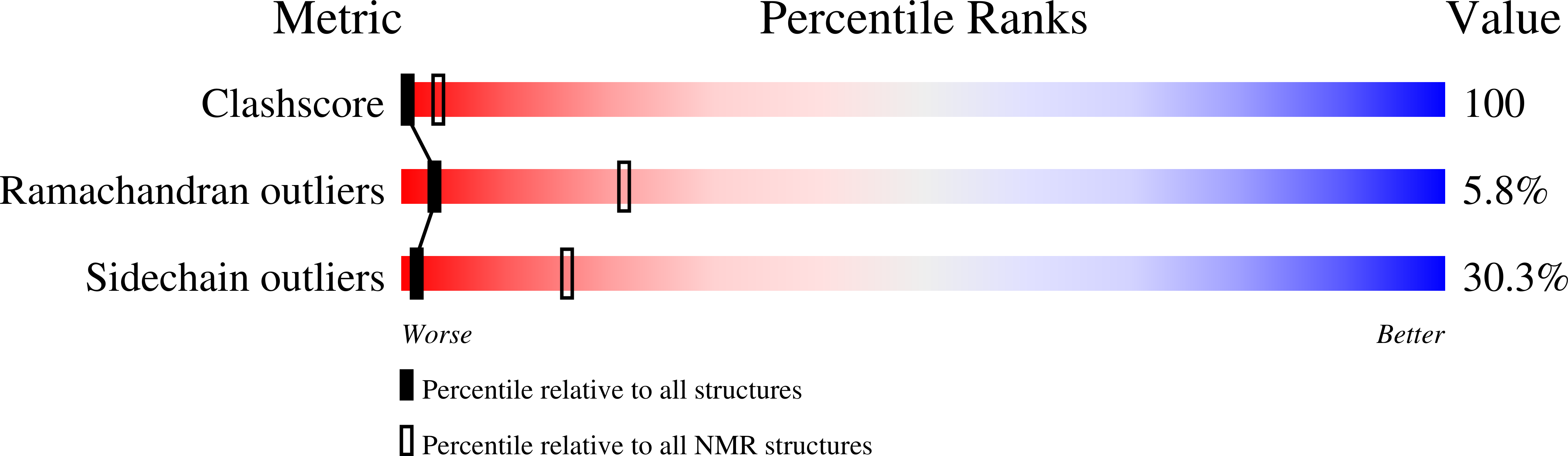
Legend

Protein

Chemical

Disease

Primary Citation of related structures