Deposition Date
2004-03-19
Release Date
2004-06-15
Last Version Date
2023-08-23
Entry Detail
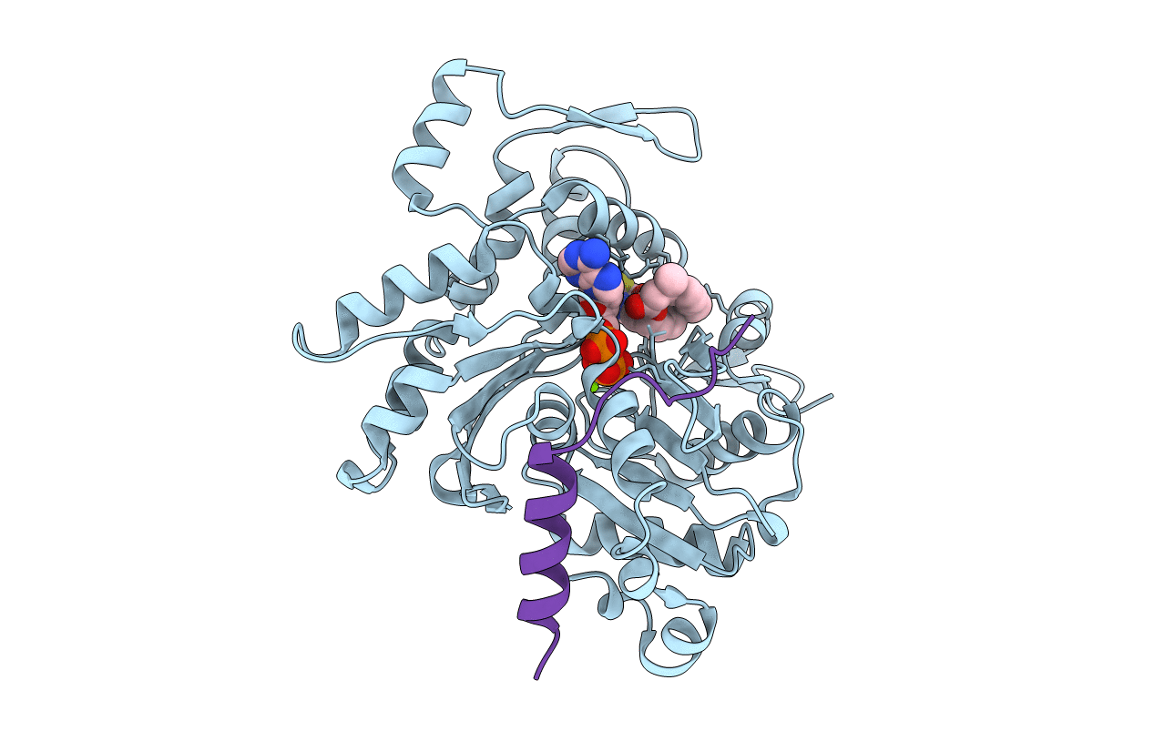
PDB ID:
1SQK
Keywords:
Title:
CRYSTAL STRUCTURE OF CIBOULOT IN COMPLEX WITH SKELETAL ACTIN
Biological Source:
Source Organism(s):
Drosophila melanogaster (Taxon ID: 7227)
Oryctolagus cuniculus (Taxon ID: 9986)
Oryctolagus cuniculus (Taxon ID: 9986)
Expression System(s):
Method Details:
Experimental Method:
Resolution:
2.50 Å
R-Value Free:
0.27
R-Value Work:
0.21
Space Group:
P 21 21 21