Deposition Date
1997-09-01
Release Date
1997-12-17
Last Version Date
2024-02-14
Entry Detail
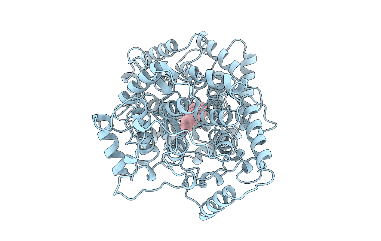
PDB ID:
1SQC
Keywords:
Title:
SQUALENE-HOPENE-CYCLASE FROM ALICYCLOBACILLUS ACIDOCALDARIUS
Biological Source:
Source Organism(s):
Alicyclobacillus acidocaldarius (Taxon ID: 405212)
Expression System(s):
Method Details:
Experimental Method:
Resolution:
2.85 Å
R-Value Free:
0.24
R-Value Work:
0.16
R-Value Observed:
0.16
Space Group:
P 3 2 1