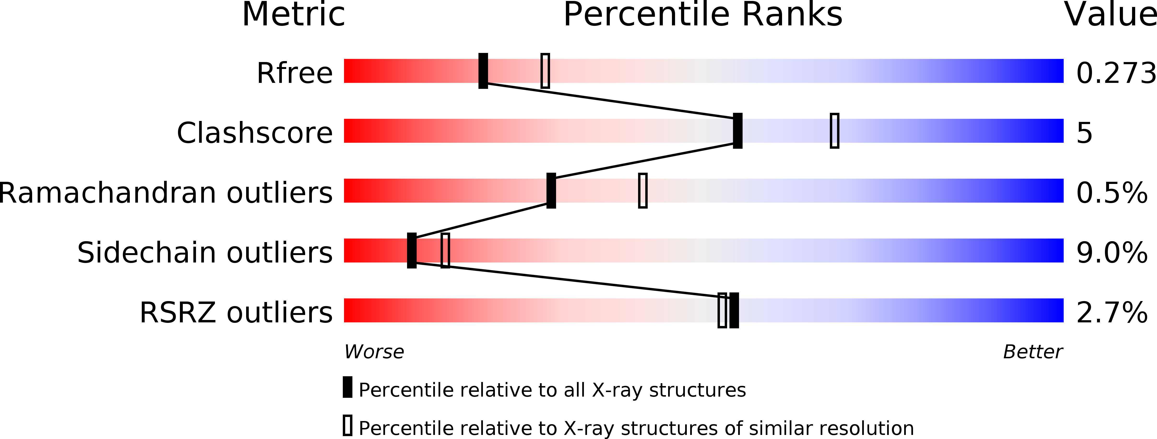
1SQ6 image
Deposition Date 2004-03-17
Release Date 2004-04-06
Last Version Date 2024-11-20
Entry Detail
PDB ID:
1SQ6
Keywords:
Title:
Plasmodium falciparum homolog of Uridine phosphorylase/Purine nucleoside phosphorylase
Biological Source:
Source Organism:
Host Organism:
Method Details:
Experimental Method:
Resolution:
2.40 Å
R-Value Free:
0.26
R-Value Work:
0.18
R-Value Observed:
0.18
Space Group:
H 3 2
Macromolecular Entities
Polymer Type:polypeptide(L)
Molecule:uridine phosphorylase, putative
Gene (Uniprot):PNP
Chain IDs:A
Chain Length:253
Number of Molecules:1
Biological Source:Plasmodium falciparum
Modified Residue
Compound ID Chain ID Parent Comp ID Details 2D Image
MSE A MET SELENOMETHIONINE
Ligand Molecules
Primary Citation
Structures of Plasmodium falciparum purine nucleoside phosphorylase complexed with sulfate and its natural substrate inosine.
Acta Crystallogr.,Sect.D 61 1245 1254 (2005)
PMID: 16131758 DOI: 10.1107/S0907444905020251

Abstact

Purine metabolism in the parasite Plasmodium has been identified as a promising target for antimalarial therapies. Purine nucleoside phosphorylase (PNP) is part of a salvage pathway for the biosynthesis of purines, which are essential for parasite survival. Two crystal structures of PNP from Plasmodium falciparum (PfPNP) in two space groups, each with a single subunit in the asymmetric unit, are described here. One structure, refined to 2.4 A, has an empty nucleoside-binding site and a sulfate ion bound in the phosphate-binding pocket. The second structure, refined to 2.0 A, has the substrate inosine bound to the active centre. Structure comparison reveals alterations in the active site upon ligand binding. The new structures presented here specifically highlight the likely roles of Asp206 and two loops flanking the active site: the beta7-alpha6 loop (residues approximately 161-169) and the beta9-alpha8 loop (residues approximately 208-223). Comparison with PNP in complex with transition-state inhibitors suggests that the purine substrate moves towards the phosphate substrate, rather than vice versa, upon forming the transition state. The single-substrate-containing PfPNP structures also appear to be more flexible than PfPNP bound to inhibitors. Together, these structures serve as a basis for better understanding of ligand binding and mechanism that can be further exploited to optimize the specificity of anti-PfPNP drugs.

Legend

Protein

Chemical

Disease

Primary Citation of related structures