Deposition Date
2004-03-17
Release Date
2004-04-13
Last Version Date
2024-11-13
Entry Detail
PDB ID:
1SQ0
Keywords:
Title:
Crystal Structure of the Complex of the Wild-type Von Willebrand Factor A1 domain and Glycoprotein Ib alpha at 2.6 Angstrom Resolution
Biological Source:
Source Organism(s):
Homo sapiens (Taxon ID: 9606)
Expression System(s):
Method Details:
Experimental Method:
Resolution:
2.60 Å
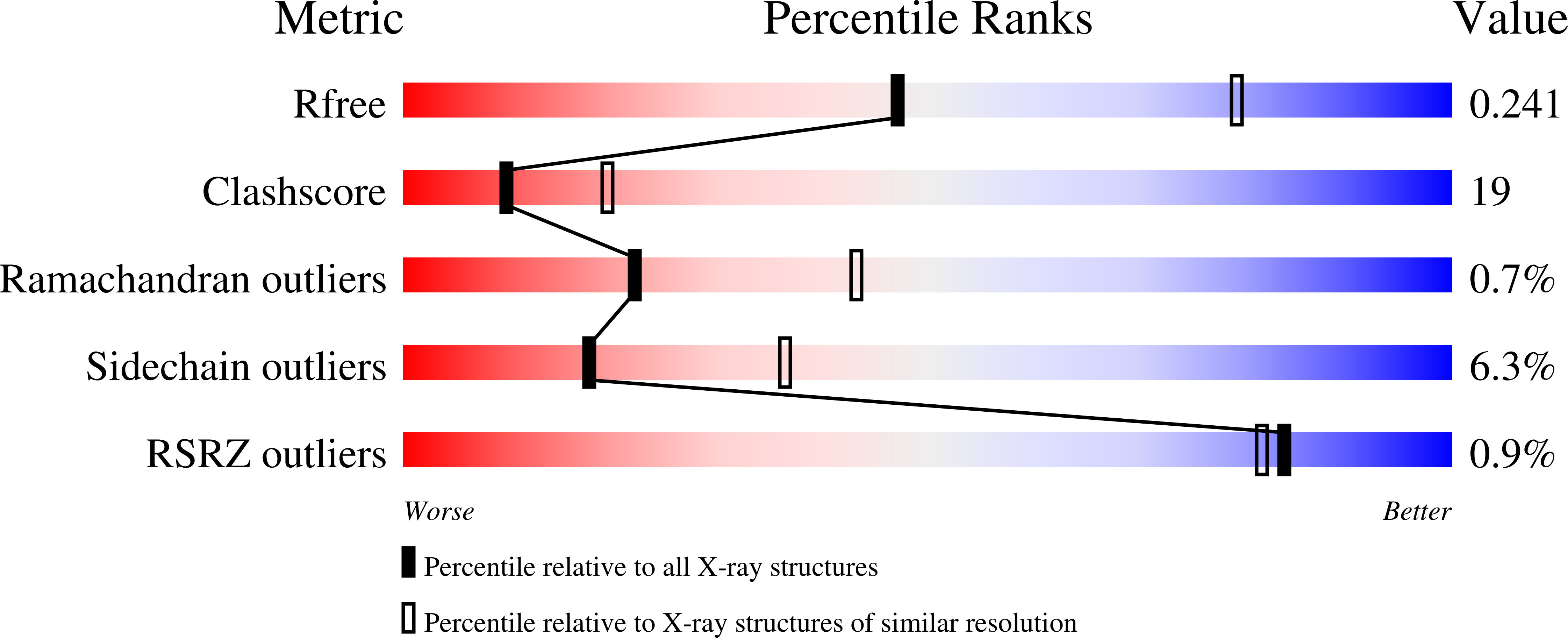
R-Value Free:
0.23
R-Value Work:
0.19
R-Value Observed:
0.19
Space Group:
P 21 21 2