Deposition Date
2004-03-15
Release Date
2004-05-11
Last Version Date
2023-08-23
Entry Detail
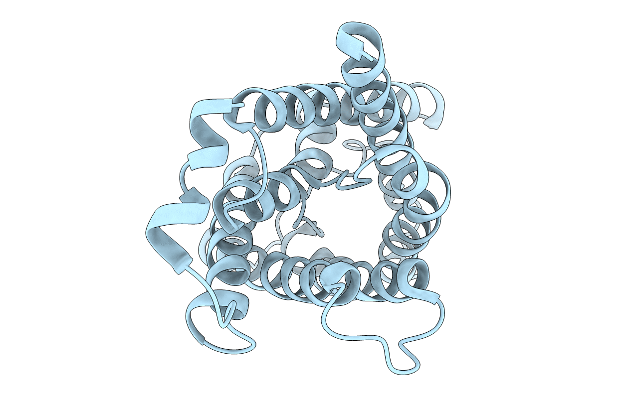
PDB ID:
1SOR
Keywords:
Title:
Aquaporin-0 membrane junctions reveal the structure of a closed water pore
Biological Source:
Source Organism(s):
Ovis aries (Taxon ID: 9940)
Method Details:
Experimental Method:
Resolution:
3.00 Å
R-Value Free:
0.33
R-Value Work:
0.29
R-Value Observed:
0.29
Space Group:
P 4 2 2