1SOI image
Deposition Date 2004-03-15
Release Date 2004-05-11
Last Version Date 2024-02-14
Entry Detail
PDB ID:
1SOI
Keywords:
Title:
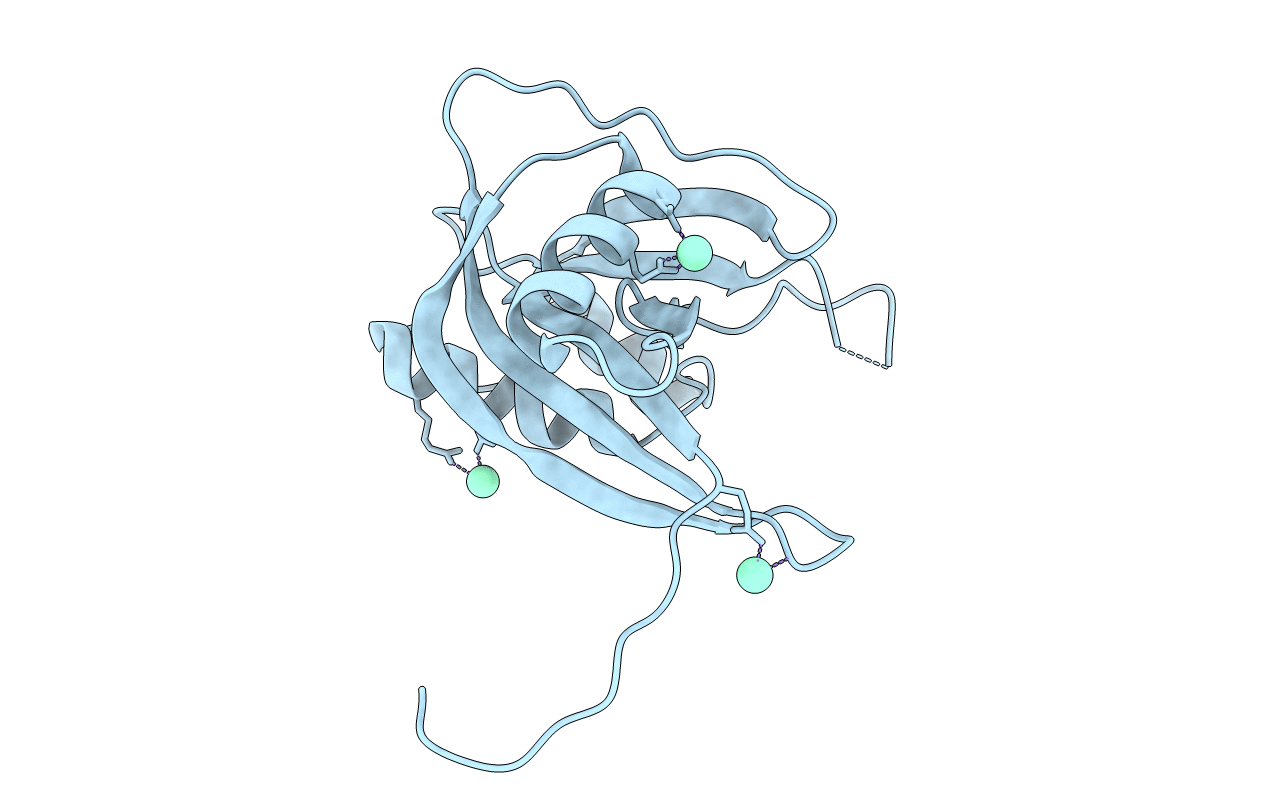
CRYSTAL STRUCTURE OF NUDIX HYDROLASE DR1025 IN COMPLEX WITH SM+3
Biological Source:
Source Organism:
Host Organism:
Method Details:
Experimental Method:
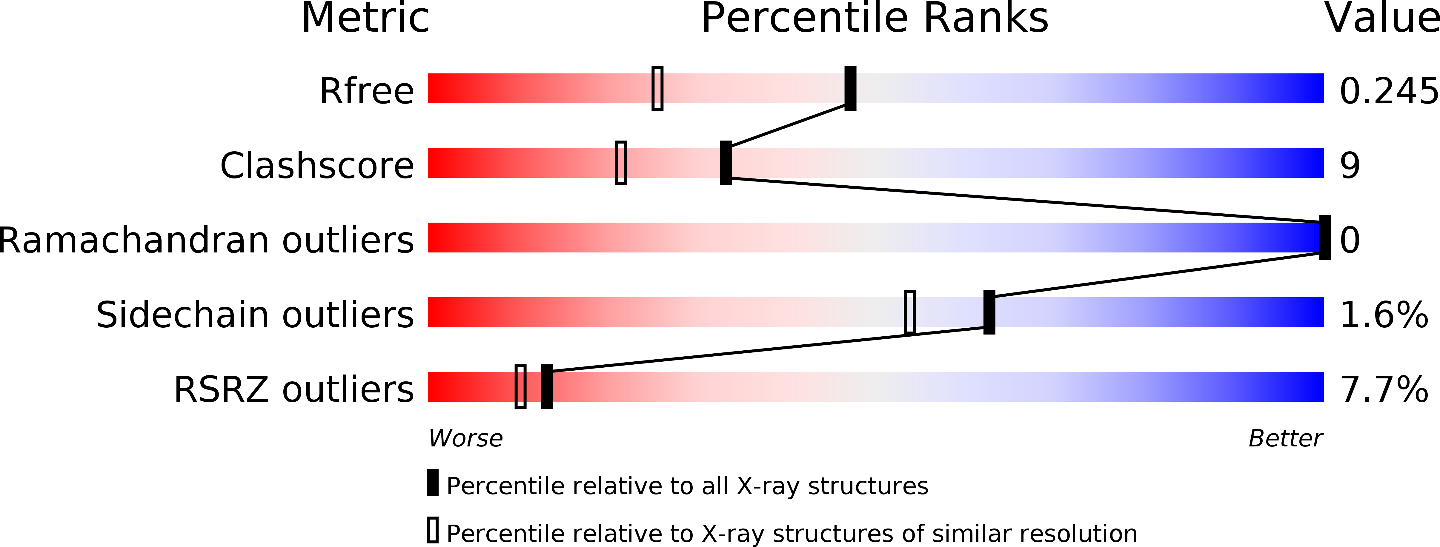
Resolution:
1.80 Å
R-Value Free:
0.25
R-Value Work:
0.21
R-Value Observed:
0.21
Space Group:
P 41 21 2
Macromolecular Entities
Polymer Type:polypeptide(L)
Molecule:MutT/nudix family protein
Gene (Uniprot):DR_1025
Chain IDs:A
Chain Length:159
Number of Molecules:1
Biological Source:Deinococcus radiodurans
Ligand Molecules
Primary Citation
Structural Studies of the Nudix Hydrolase DR1025 From Deinococcus radiodurans and its Ligand Complexes.
J.Mol.Biol. 339 103 116 (2004)
PMID: 15123424 DOI: 10.1016/j.jmb.2004.01.065

Abstact

We have determined the crystal structure, at 1.4A, of the Nudix hydrolase DR1025 from the extremely radiation resistant bacterium Deinococcus radiodurans. The protein forms an intertwined homodimer by exchanging N-terminal segments between chains. We have identified additional conserved elements of the Nudix fold, including the metal-binding motif, a kinked beta-strand characterized by a proline two positions upstream of the Nudix consensus sequence, and participation of the N-terminal extension in the formation of the substrate-binding pocket. Crystal structures were also solved of DR1025 crystallized in the presence of magnesium and either a GTP analog or Ap(4)A (both at 1.6A resolution). In the Ap(4)A co-crystal, the electron density indicated that the product of asymmetric hydrolysis, ATP, was bound to the enzyme. The GTP analog bound structure showed that GTP was bound almost identically as ATP. Neither nucleoside triphosphate was further cleaved.

Legend

Protein

Chemical

Disease

Primary Citation of related structures