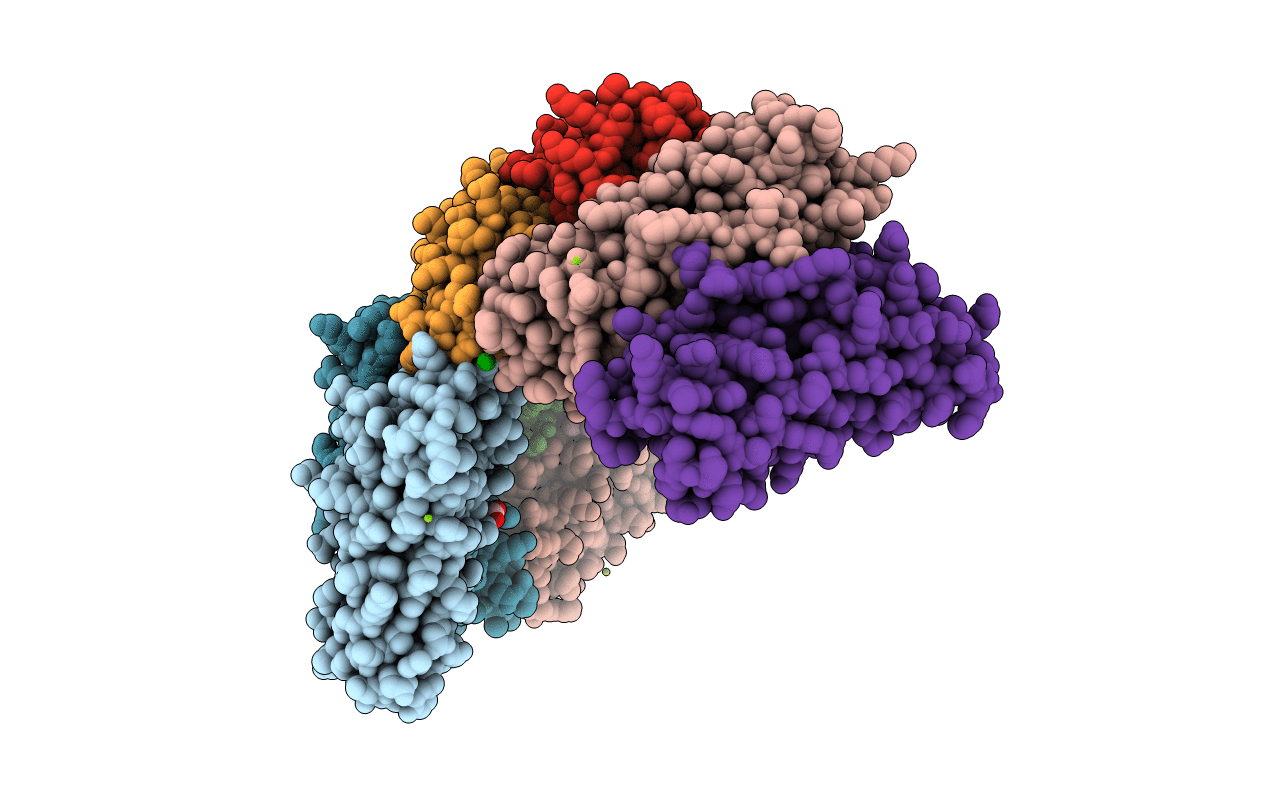
1SOF image
Entry Detail
PDB ID:
1SOF
Title:
Crystal structure of the azotobacter vinelandii bacterioferritin at 2.6 A resolution
Biological Source:
Source Organism:
PDB Version:
Deposition Date:
2004-03-14
Release Date:
2005-04-05
Method Details:
Experimental Method:
Resolution:
2.60 Å
R-Value Free:
0.24
R-Value Work:
0.19
R-Value Observed:
0.19
Space Group:
H 3
Macromolecular Entities
Polymer Type:polypeptide(L)
Description:Bacterioferritin
Chain IDs:A, B, C, D, E, F, G, H
Chain Length:156
Number of Molecules:8
Biological Source:Azotobacter vinelandii
Primary Citation
2.6 A resolution crystal structure of the bacterioferritin from Azotobacter vinelandii
Febs Lett. 573 93 98 (2004)
PMID: 15327981 DOI: 10.1016/j.febslet.2004.07.054

Abstact

The crystal structure of the bacterioferritin from Azotobacter vinelandii has been determined at 2.6 A resolution. Both the low occupancy of one iron ion in the dinuclear iron center and the deviation of its adjacent residue His130 from the center suggest migration of the iron ion from the dinuclear iron site to the inner nucleation site. The concerted movement of His130 and Glu47 may admit a dynamic gating mechanism for shift of the oxidized iron ion. Ba2+ binding to the fourfold channel implicates that the channel bears Fe2+ conductivity and selectivity to provide a route for iron access to the inner cavity during core formation.

Legend

Protein

Chemical

Disease

Primary Citation of related structures