1SO7 image
Deposition Date 2004-03-12
Release Date 2004-11-02
Last Version Date 2024-11-06
Entry Detail
PDB ID:
1SO7
Keywords:
Title:
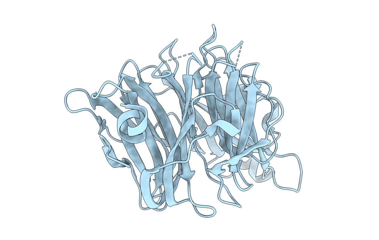
Maltose-induced structure of the human cytolsolic sialidase Neu2
Biological Source:
Source Organism:
Homo sapiens (Taxon ID: 9606)
Host Organism:
Method Details:
Experimental Method:
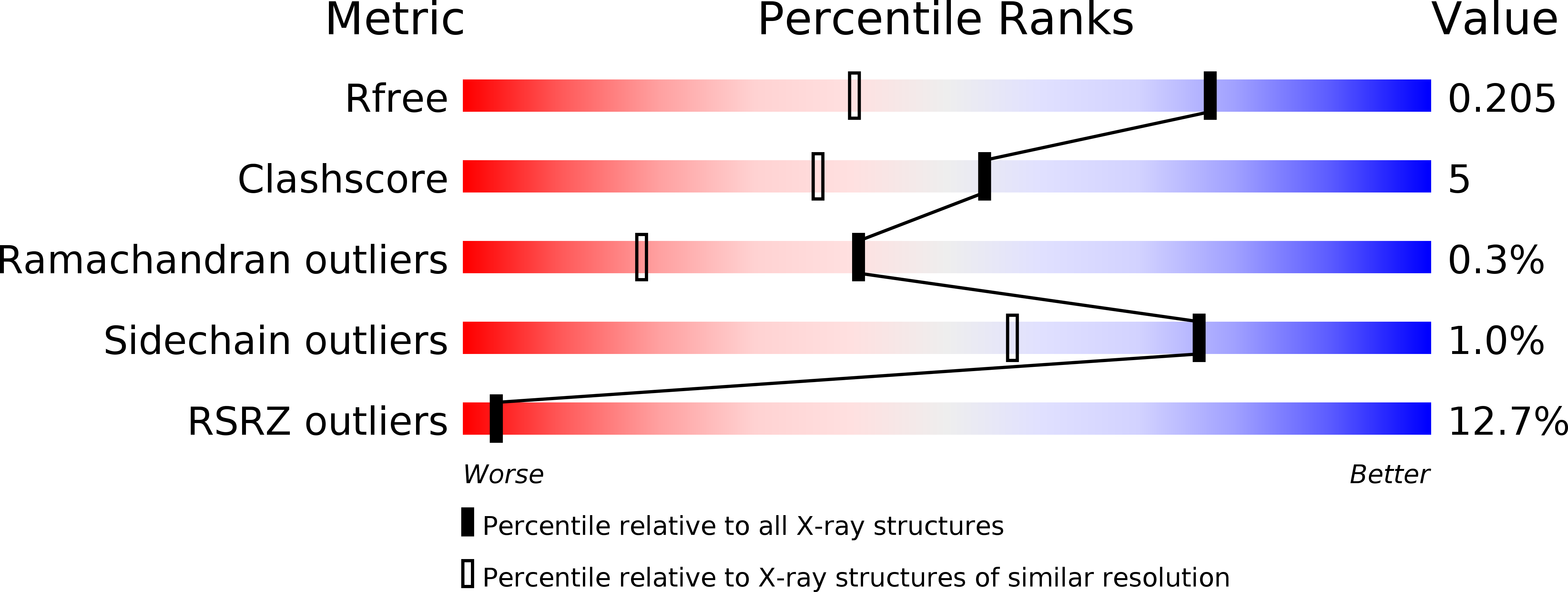
Resolution:
1.49 Å
R-Value Free:
0.21
R-Value Work:
0.20
R-Value Observed:
0.20
Space Group:
H 3
Macromolecular Entities
Structures with similar UniProt ID
Protein Blast
Polymer Type:polypeptide(L)
Molecule:Sialidase 2
Gene (Uniprot):NEU2
Chain IDs:A
Chain Length:382
Number of Molecules:1
Biological Source:Homo sapiens
Ligand Molecules
Primary Citation
Crystal Structure of the Human Cytosolic Sialidase Neu2: EVIDENCE FOR THE DYNAMIC NATURE OF SUBSTRATE RECOGNITION
J.Biol.Chem. 280 469 475 (2005)
PMID: 15501818 DOI: 10.1074/jbc.M411506200

Abstact

Gangliosides play key roles in cell differentiation, cell-cell interactions, and transmembrane signaling. Sialidases hydrolyze sialic acids to produce asialo compounds, which is the first step of degradation processes of glycoproteins and gangliosides. Sialidase involvement has been implicated in some lysosomal storage disorders such as sialidosis and galactosialidosis. Neu2 is a recently identified human cytosolic sialidase. Here we report the first high resolution x-ray structures of mammalian sialidase, human Neu2, in its apo form and in complex with an inhibitor, 2-deoxy-2,3-dehydro-N-acetylneuraminic acid (DANA). The structure shows the canonical six-blade beta-propeller observed in viral and bacterial sialidases with its active site in a shallow crevice. In the complex structure, the inhibitor lies in the catalytic crevice surrounded by ten amino acids. In particular, the arginine triad, conserved among sialidases, aids in the proper positioning of the carboxylate group of DANA within the active site region. The tyrosine residue, Tyr(334), conserved among mammalian and bacterial sialidases as well as in viral neuraminidases, facilitates the enzymatic reaction by stabilizing a putative carbonium ion in the transition state. The loops containing Glu(111) and the catalytic aspartate Asp(46) are disordered in the apo form but upon binding of DANA become ordered to adopt two short alpha-helices to cover the inhibitor, illustrating the dynamic nature of substrate recognition. The N-acetyl and glycerol moieties of DANA are recognized by Neu2 residues not shared by bacterial sialidases and viral neuraminidases, which can be regarded as a key structural difference for potential drug design against bacteria, influenza, and other viruses.

Legend

Protein

Chemical

Disease

Primary Citation of related structures
Feedback Form
Name
Email
Institute
Feedback