
Deposition Date
2004-03-09
Release Date
2004-03-16
Last Version Date
2024-04-03
Entry Detail
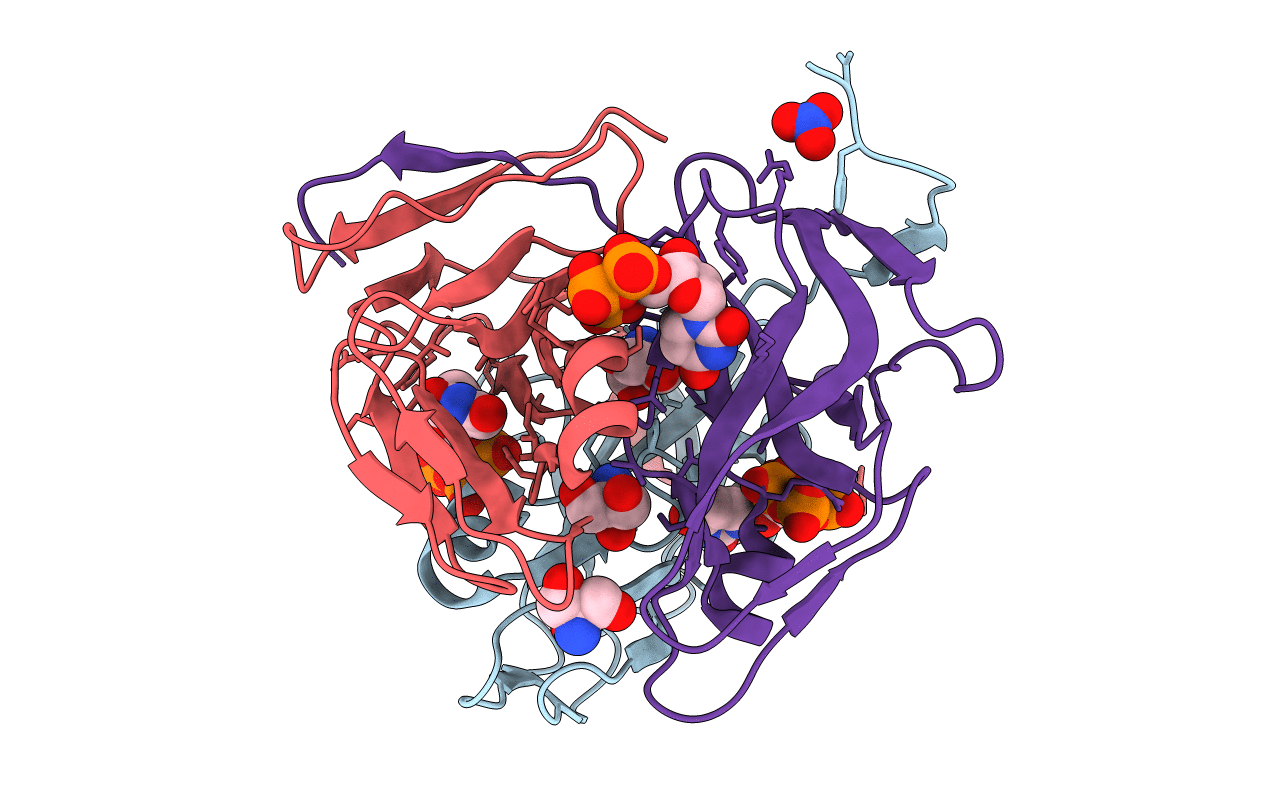
PDB ID:
1SMC
Keywords:
Title:
Mycobacterium tuberculosis dUTPase complexed with dUTP in the absence of metal ion.
Biological Source:
Source Organism(s):
Mycobacterium tuberculosis (Taxon ID: 1773)
Expression System(s):
Method Details:
Experimental Method:
Resolution:
2.10 Å
R-Value Free:
0.20
R-Value Work:
0.16
R-Value Observed:
0.16
Space Group:
P 21 21 21
