1SL4 image
Entry Detail
PDB ID:
1SL4
Title:
Crystal Structure of DC-SIGN carbohydrate recognition domain complexed with Man4
Biological Source:
Source Organism:
Host Organism:
PDB Version:
Deposition Date:
2004-03-05
Release Date:
2004-06-15
Method Details:
Experimental Method:
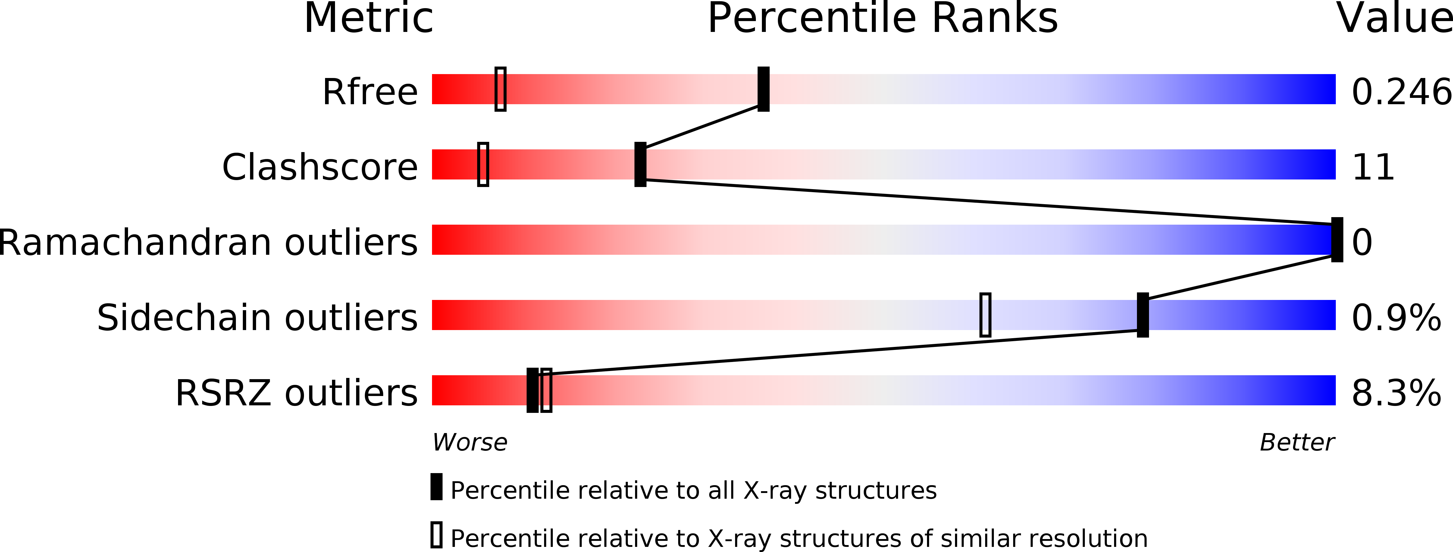
Resolution:
1.55 Å
R-Value Free:
0.25
R-Value Work:
0.22
Space Group:
P 43
Macromolecular Entities
Polymer Type:polypeptide(L)
Description:mDC-SIGN1B type I isoform
Chain IDs:A
Chain Length:155
Number of Molecules:1
Biological Source:Homo sapiens
Ligand Molecules
Primary Citation
Structural basis for distinct ligand-binding and targeting properties of the receptors DC-SIGN and DC-SIGNR
Nat.Struct.Mol.Biol. 11 591 598 (2004)
PMID: 15195147 DOI: 10.1038/nsmb784

Abstact

Both the dendritic cell receptor DC-SIGN and the closely related endothelial cell receptor DC-SIGNR bind human immunodeficiency virus and enhance infection. However, biochemical and structural comparison of these receptors now reveals that they have very different physiological functions. By screening an extensive glycan array, we demonstrated that DC-SIGN and DC-SIGNR have distinct ligand-binding properties. Our structural and mutagenesis data explain how both receptors bind high-mannose oligosaccharides on enveloped viruses and why only DC-SIGN binds blood group antigens, including those present on microorganisms. DC-SIGN mediates endocytosis, trafficking as a recycling receptor and releasing ligand at endosomal pH, whereas DC-SIGNR does not release ligand at low pH or mediate endocytosis. Thus, whereas DC-SIGN has dual ligand-binding properties and functions both in adhesion and in endocytosis of pathogens, DC-SIGNR binds a restricted set of ligands and has only the properties of an adhesion receptor.

Legend

Protein

Chemical

Disease

Primary Citation of related structures