1SL0 image
Entry Detail
PDB ID:
1SL0
Title:
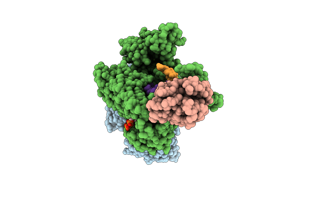
Ternary 3' complex of T7 DNA polymerase with a DNA primer/template containing a disordered cis-syn thymine dimer on the template and an incoming nucleotide
Biological Source:
Host Organism:
PDB Version:
Deposition Date:
2004-03-05
Release Date:
2004-07-06
Method Details:
Experimental Method:
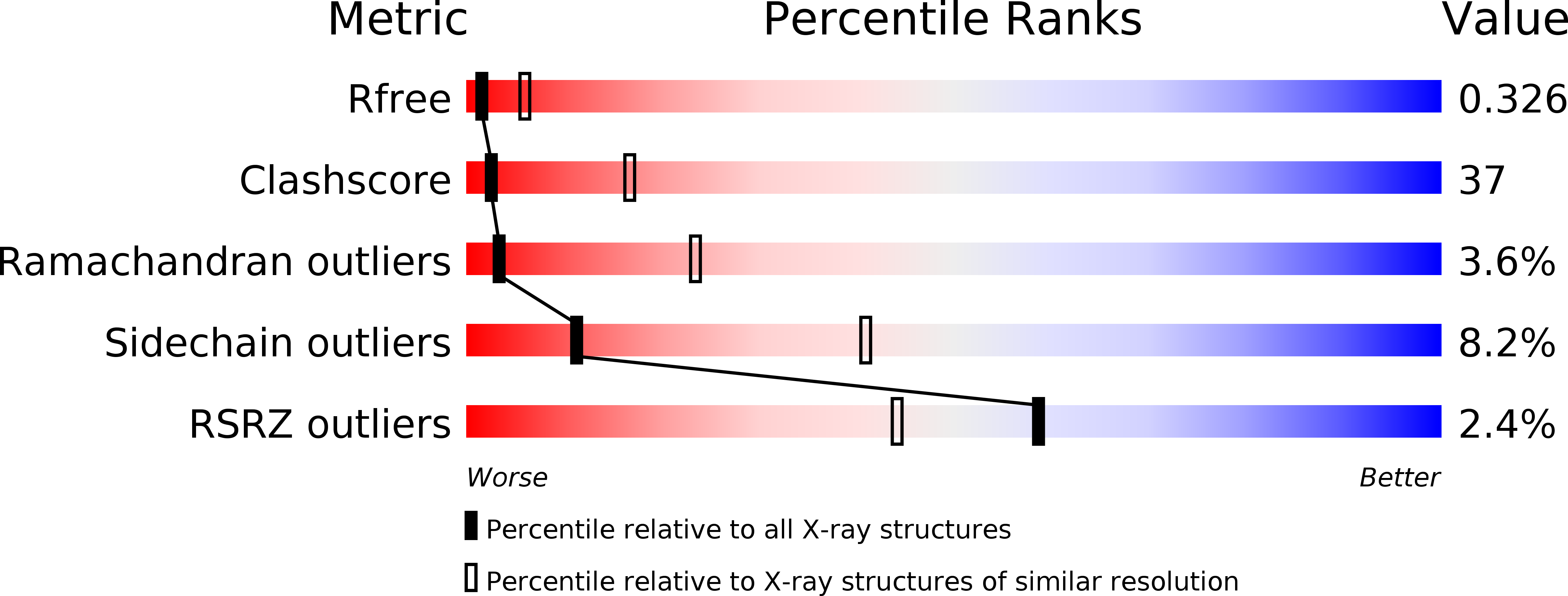
Resolution:
3.20 Å
R-Value Free:
0.35
R-Value Work:
0.28
R-Value Observed:
0.28
Space Group:
P 1 21 1
Macromolecular Entities
Polymer Type:polypeptide(L)
Description:DNA polymerase
Mutations:DEL(118-123)
Chain IDs:E (auth: A), G (auth: C)
Chain Length:698
Number of Molecules:2
Biological Source:Enterobacteria phage T7
Polymer Type:polypeptide(L)
Description:Thioredoxin 1
Chain IDs:F (auth: B), H (auth: D)
Chain Length:108
Number of Molecules:2
Biological Source:Escherichia coli
Modified Residue
Compound ID Chain ID Parent Comp ID Details 2D Image
2DT A DT 3'-DEOXYTHYMIDINE-5'-MONOPHOSPHATE
Primary Citation
Nucleotide insertion opposite a cis-syn thymine dimer by a replicative DNA polymerase from bacteriophage T7.
Nat.Struct.Mol.Biol. 11 784 790 (2004)
PMID: 15235589 DOI: 10.1038/nsmb792

Abstact

Ultraviolet-induced DNA damage poses a lethal block to replication. To understand the structural basis for this, we determined crystal structures of a replicative DNA polymerase from bacteriophage T7 in complex with nucleotide substrates and a DNA template containing a cis-syn cyclobutane pyrimidine dimer (CPD). When the 3' thymine is the templating base, the CPD is rotated out of the polymerase active site and the fingers subdomain adopts an open orientation. When the 5' thymine is the templating base, the CPD lies within the polymerase active site where it base-pairs with the incoming nucleotide and the 3' base of the primer, while the fingers are in a closed conformation. These structures reveal the basis for the strong block of DNA replication that is caused by this photolesion.

Legend

Protein

Chemical

Disease

Primary Citation of related structures