Deposition Date
2004-03-04
Release Date
2004-07-13
Last Version Date
2024-11-13
Entry Detail
PDB ID:
1SK4
Keywords:
Title:
crystal structure of the C-terminal peptidoglycan-binding domain of human peptidoglycan recognition protein Ialpha
Biological Source:
Source Organism:
Homo sapiens (Taxon ID: 9606)
Host Organism:
Method Details:
Experimental Method:
Resolution:
1.65 Å
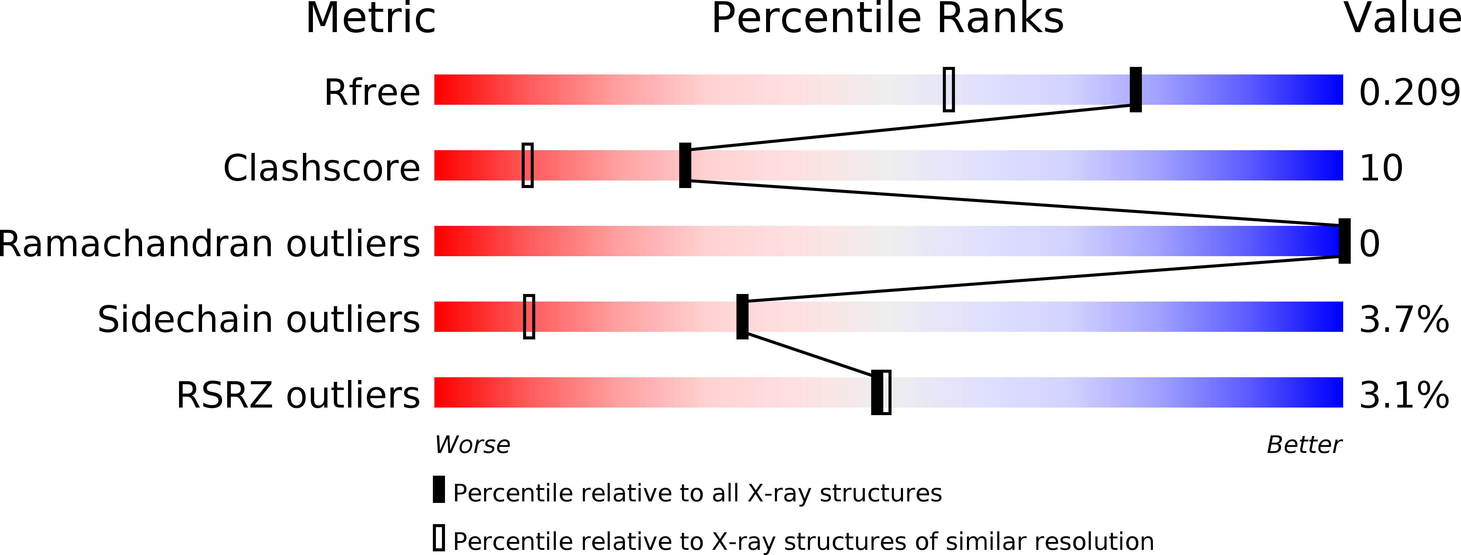
R-Value Free:
0.21
R-Value Work:
0.19
R-Value Observed:
0.19
Space Group:
P 32 2 1