
Deposition Date
2004-03-03
Release Date
2005-03-15
Last Version Date
2024-02-14
Entry Detail
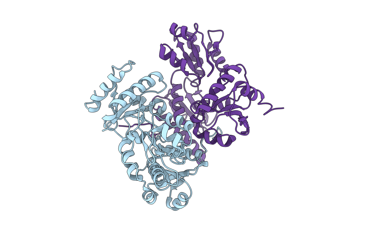
PDB ID:
1SJI
Keywords:
Title:
Comparing skeletal and cardiac calsequestrin structures and their calcium binding: a proposed mechanism for coupled calcium binding and protein polymerization
Biological Source:
Source Organism:
Canis lupus familiaris (Taxon ID: 9615)
Host Organism:
Method Details:
Experimental Method:
Resolution:
2.40 Å
R-Value Free:
0.24
R-Value Work:
0.19
R-Value Observed:
0.19
Space Group:
I 4
