1SJ9 image
Deposition Date 2004-03-03
Release Date 2005-03-08
Last Version Date 2023-08-23
Entry Detail
PDB ID:
1SJ9
Keywords:
Title:
Crystal structure of the uridine phosphorylase from Salmonella typhimurium at 2.5A resolution
Biological Source:
Source Organism:
Method Details:
Experimental Method:
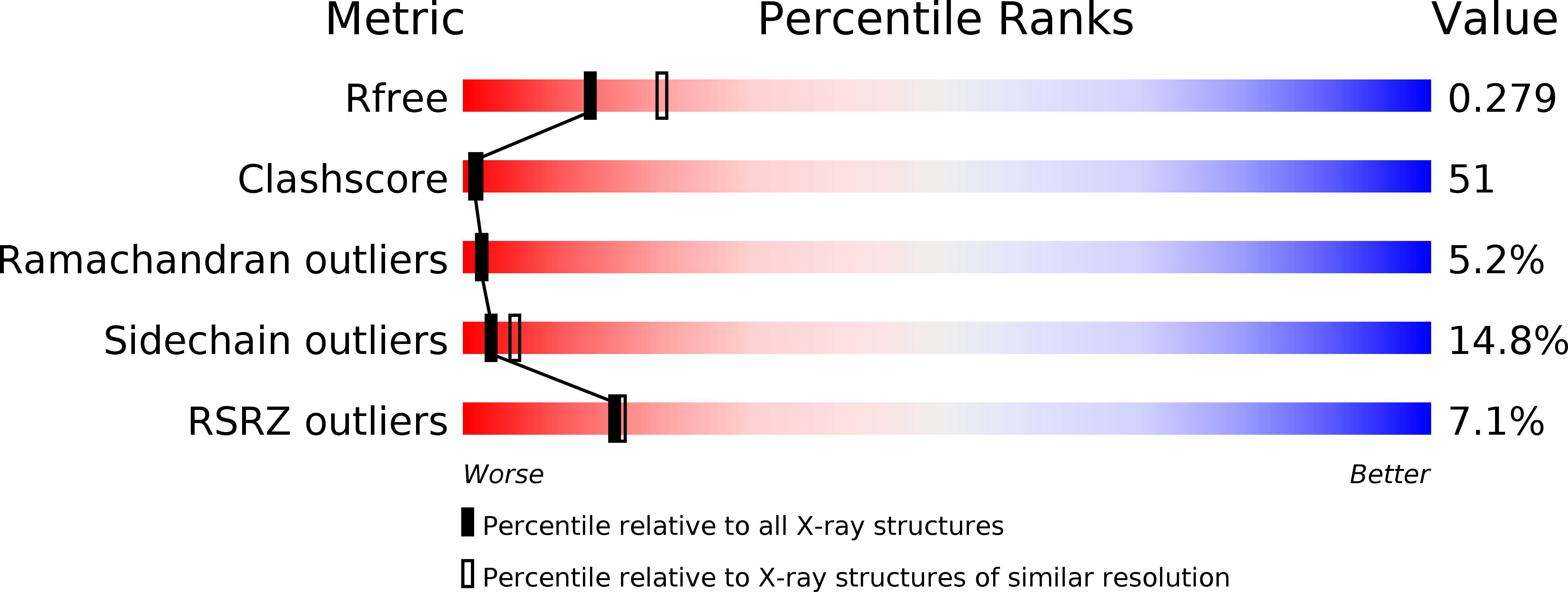
Resolution:
2.50 Å
R-Value Free:
0.27
R-Value Work:
0.22
R-Value Observed:
0.22
Space Group:
P 61
Macromolecular Entities
Structures with similar UniProt ID
Protein Blast
Polymer Type:polypeptide(L)
Molecule:Uridine phosphorylase
Gene (Uniprot):udp
Chain IDs:A, B, C, D, E, F
Chain Length:253
Number of Molecules:6
Biological Source:Salmonella typhimurium
Ligand Molecules
Primary Citation
Preliminary investigation of the three-dimensional structure of Salmonella typhimurium uridine phosphorylase in the crystalline state.
Acta Crystallogr.,Sect.F 61 337 340 (2005)
PMID: 16511035 DOI: 10.1107/S1744309105007463

Abstact

Uridine phosphorylase (UPh) catalyzes the phosphorolytic cleavage of the C-N glycosidic bond of uridine to ribose 1-phosphate and uracil in the pyrimidine-salvage pathway. The crystal structure of the Salmonella typhimurium uridine phosphorylase (StUPh) has been determined at 2.5 A resolution and refined to an R factor of 22.1% and an Rfree of 27.9%. The hexameric StUPh displays 32 point-group symmetry and utilizes both twofold and threefold non-crystallographic axes. A phosphate is bound at the active site and forms hydrogen bonds to Arg91, Arg30, Thr94 and Gly26 of one monomer and Arg48 of an adjacent monomer. The hexameric StUPh model reveals a close structural relationship to Escherichia coli uridine phosphorylase (EcUPh).

Legend

Protein

Chemical

Disease

Primary Citation of related structures