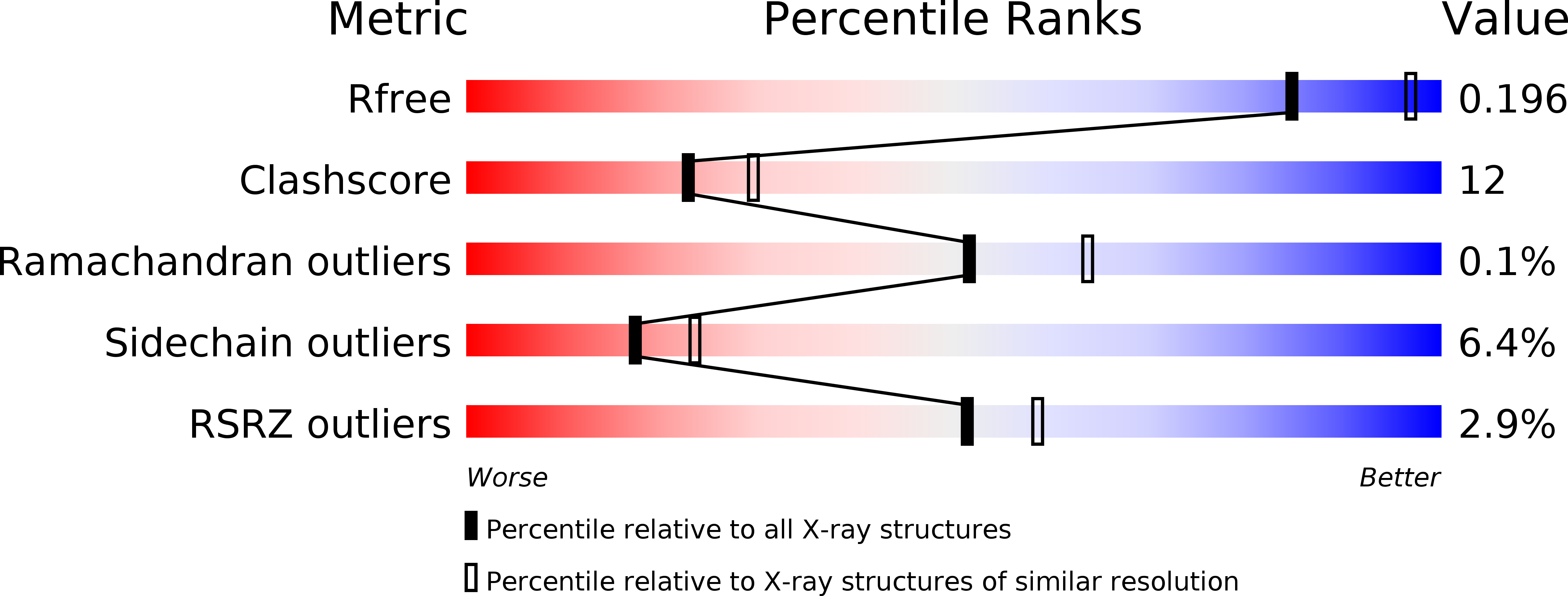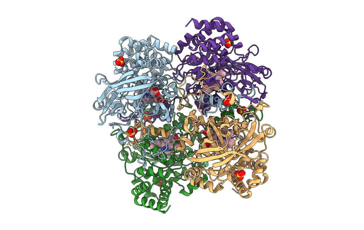
1SI8 image
Deposition Date 2004-02-28
Release Date 2004-05-04
Last Version Date 2023-08-23
Entry Detail
PDB ID:
1SI8
Keywords:
Title:
Crystal structure of E. faecalis catalase
Biological Source:
Source Organism:
Host Organism:
Method Details:
Experimental Method:
Resolution:
2.30 Å
R-Value Free:
0.21
R-Value Work:
0.20
R-Value Observed:
0.20
Space Group:
H 3
Macromolecular Entities
Structures with similar UniProt ID
Protein Blast
Polymer Type:polypeptide(L)
Molecule:Catalase
Gene (Uniprot):katA
Chain IDs:A, B, C, D
Chain Length:484
Number of Molecules:4
Biological Source:Enterococcus faecalis
Primary Citation
The three-dimensional structure of catalase from Enterococcus faecalis.
Acta Crystallogr.,Sect.D 60 1374 1380 (2004)
PMID: 15272159 DOI: 10.1107/S0907444904012004

Abstact

Enterococcus faecalis haem catalase was crystallized using lithium sulfate at neutral pH. The crystals belong to space group R3, with unit-cell parameters a = b = 236.9, c = 198.1 A. The three-dimensional structure was determined by molecular replacement using a subunit of the Proteus mirabilis catalase structure. It was refined against 2.3 A synchrotron data to a free R factor of 21.8%. Like other catalases, the E. faecalis catalase is a homotetramer with a fold and structure similar to those of its structurally closest relative P. mirabilis. The solvent structure in the active site is identical in the four subunits but differs from that found in other catalases. The structural consequences of the Ramachandran outlier Ser196 are discussed.

Legend

Protein

Chemical

Disease

Primary Citation of related structures