1SGH image
Deposition Date 2004-02-23
Release Date 2004-06-29
Last Version Date 2023-08-23
Entry Detail
PDB ID:
1SGH
Title:
Moesin FERM domain bound to EBP50 C-terminal peptide
Biological Source:
Source Organism:
Homo sapiens (Taxon ID: 9606)
(Taxon ID: )
Host Organism:
Method Details:
Experimental Method:
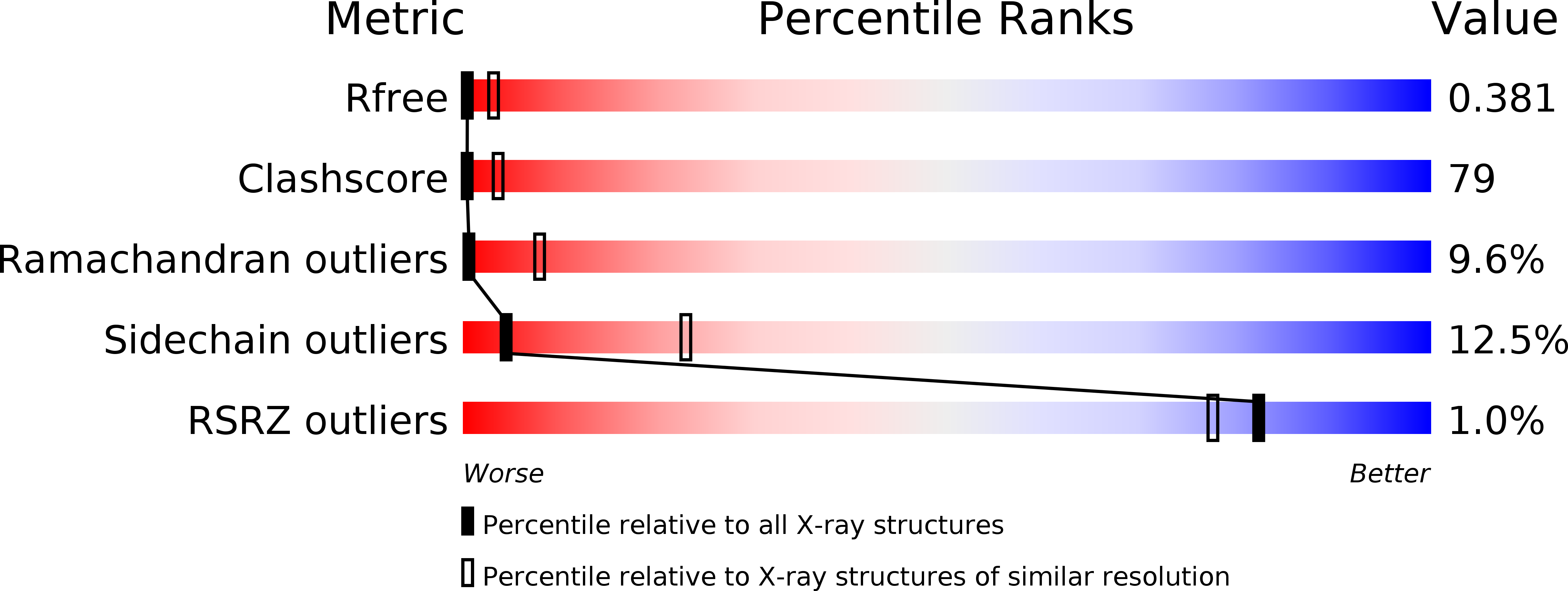
Resolution:
3.50 Å
R-Value Free:
0.40
R-Value Work:
0.33
Space Group:
C 1 2 1
Macromolecular Entities
Polymer Type:polypeptide(L)
Molecule:Moesin
Gene (Uniprot):MSN
Chain IDs:A
Chain Length:297
Number of Molecules:1
Biological Source:Homo sapiens
Polymer Type:polypeptide(L)
Molecule:Ezrin-radixin-moesin binding phosphoprotein 50
Gene (Uniprot):NHERF1
Chain IDs:B
Chain Length:39
Number of Molecules:1
Biological Source:
Ligand Molecules
Primary Citation
The EBP50-moesin interaction involves a binding site regulated by direct masking on the FERM domain
J.Cell.Sci. 117 1547 1552 (2004)
PMID: 15020681 DOI: 10.1242/jcs.01038

Abstact

Members of the ezrin-radixin-moesin (ERM) protein family serve as regulated microfilament-membrane crosslinking proteins that, upon activation, bind the scaffolding protein ERM-phosphoprotein of 50 kDa (EBP50). Here we report a 3.5 A resolution diffraction analysis of a complex between the active moesin N-terminal FERM domain and a 38 residue peptide from the C terminus of EBP50. This crystallographic result, combined with sequence and structural comparisons, suggests that the C-terminal 11 residues of EBP50 binds as an alpha-helix at the same site occupied in the dormant monomer by the last 11 residues of the inhibitory moesin C-terminal tail. Biochemical support for this interpretation derives from in vitro studies showing that appropriate mutations in both the EBP50 tail peptide and the FERM domain reduce binding, and that a peptide representing just the C-terminal 14 residues of EBP50 also binds to moesin. Combined with the recent identification of the I-CAM-2 binding site on the ERM FERM domain (Hamada, K., Shimizu, T., Yonemura, S., Tsukita, S., and Hakoshima, T. (2003) EMBO J. 22, 502-514), this study reveals that the FERM domain contains two distinct binding sites for membrane-associated proteins. The contribution of each ligand to ERM function can now be dissected by making structure-based mutations that specifically affect the binding of each ligand.

Legend

Protein

Chemical

Disease

Primary Citation of related structures