Deposition Date
2004-02-15
Release Date
2004-03-02
Last Version Date
2024-10-16
Entry Detail
PDB ID:
1SDX
Keywords:
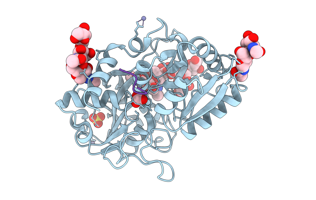
Title:
Crystal structure of the zinc saturated C-terminal half of bovine lactoferrin at 2.0 A resolution reveals two additional zinc binding sites
Biological Source:
Source Organism(s):
Bos taurus (Taxon ID: 9913)
Method Details:
Experimental Method:
Resolution:
2.06 Å
R-Value Free:
0.21
R-Value Work:
0.19
R-Value Observed:
0.19
Space Group:
P 1 21 1