
Deposition Date
2004-02-14
Release Date
2004-05-25
Last Version Date
2024-02-14
Entry Detail
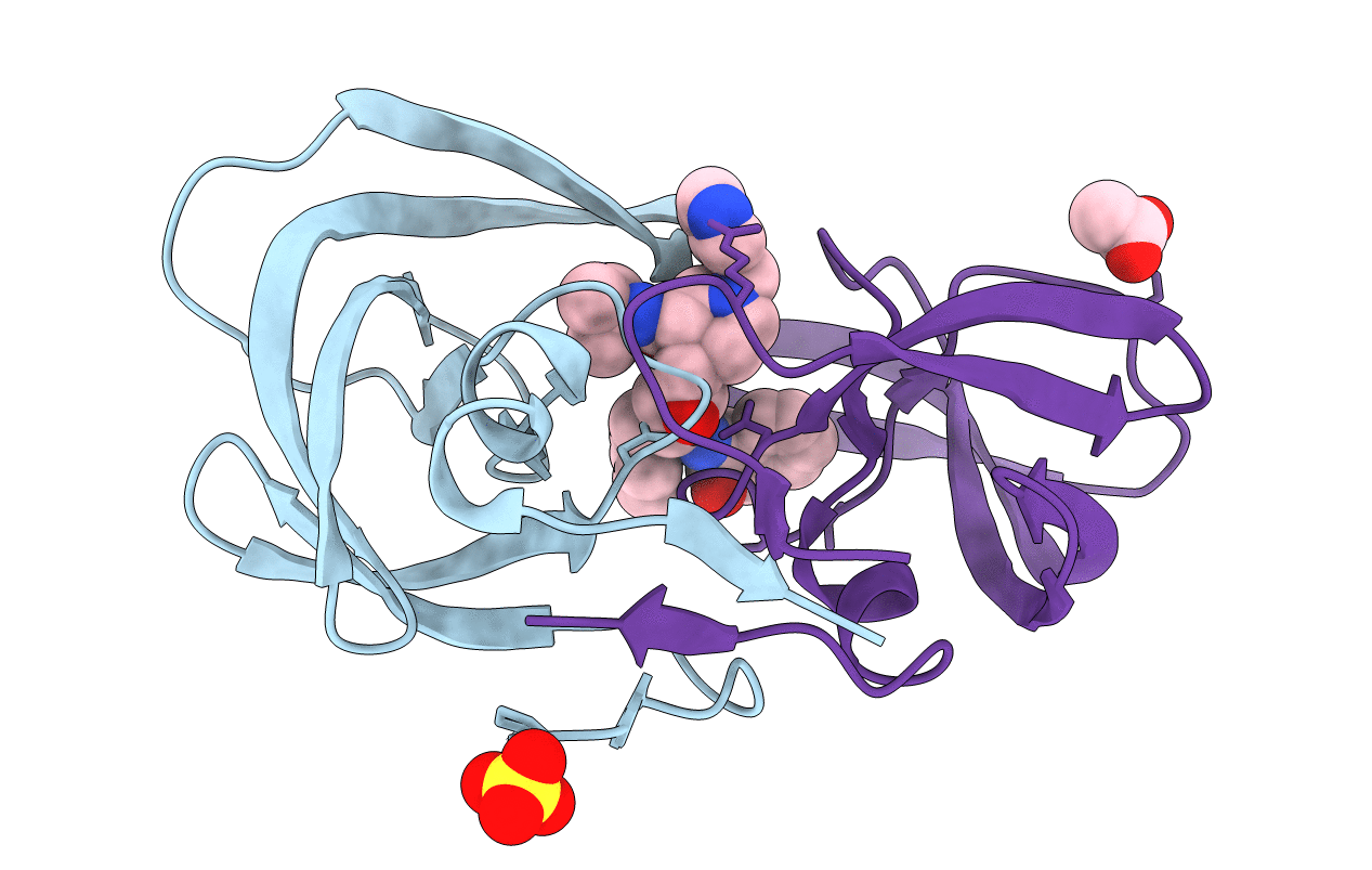
PDB ID:
1SDU
Keywords:
Title:
Crystal structures of HIV protease V82A and L90M mutants reveal changes in indinavir binding site.
Biological Source:
Source Organism(s):
Human immunodeficiency virus 1 (Taxon ID: 11676)
Expression System(s):
Method Details:
Experimental Method:
Resolution:
1.25 Å
R-Value Free:
0.18
R-Value Work:
0.14
R-Value Observed:
0.14
Space Group:
P 21 21 21
