Deposition Date
2004-02-13
Release Date
2005-02-22
Last Version Date
2023-08-23
Entry Detail
PDB ID:
1SDN
Keywords:
Title:
CRYSTAL STRUCTURE OF A DEACYLATION-DEFECTIVE MUTANT OF PENICILLIN-BINDING PROTEIN 5 MODIFIED BY MERCURY
Biological Source:
Source Organism(s):
Escherichia coli (Taxon ID: 562)
Expression System(s):
Method Details:
Experimental Method:
Resolution:
2.50 Å
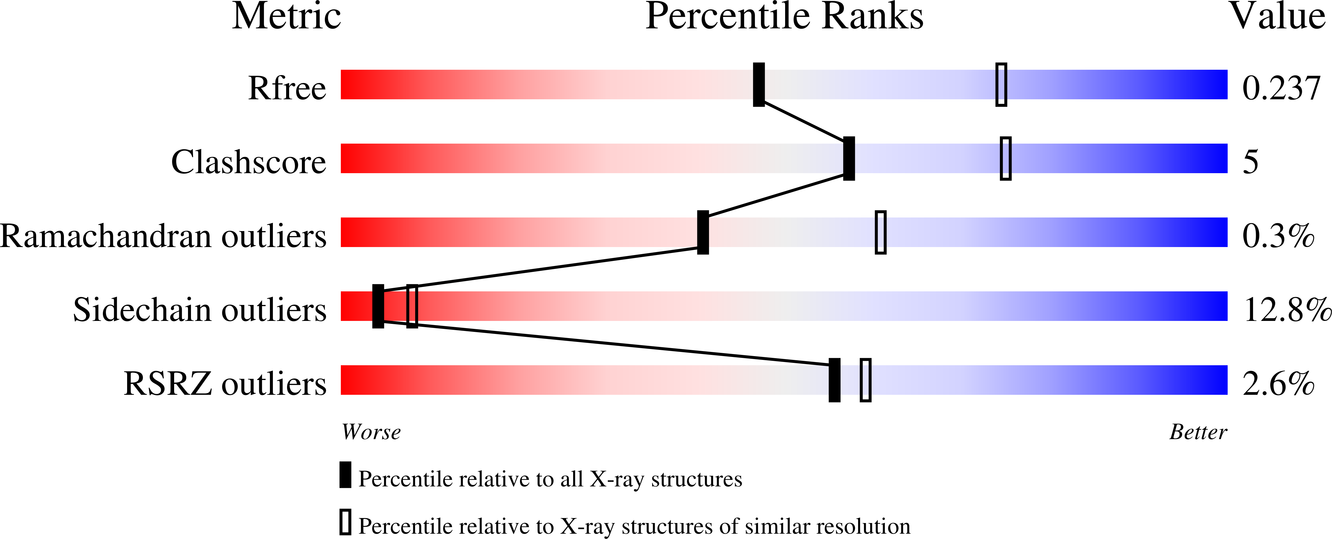
R-Value Free:
0.24
R-Value Work:
0.17
R-Value Observed:
0.17
Space Group:
P 32