1SDM image
Deposition Date 2004-02-13
Release Date 2004-06-22
Last Version Date 2023-08-23
Entry Detail
PDB ID:
1SDM
Title:
Crystal structure of kinesin-like calmodulin binding protein
Biological Source:
Source Organism:
Host Organism:
Method Details:
Experimental Method:
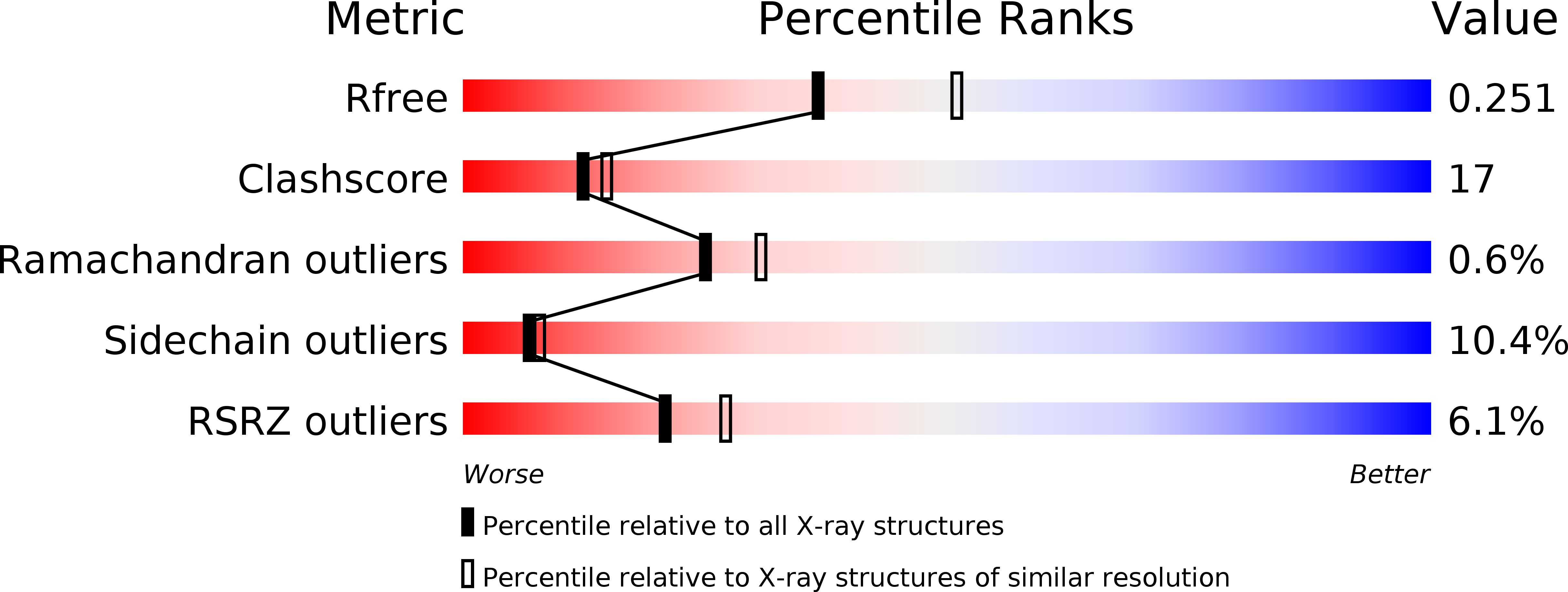
Resolution:
2.30 Å
R-Value Free:
0.25
R-Value Work:
0.21
R-Value Observed:
0.21
Space Group:
P 21 21 2
Macromolecular Entities
Polymer Type:polypeptide(L)
Molecule:kinesin heavy chain-like protein
Gene (Uniprot):LOC102577881
Chain IDs:A
Chain Length:369
Number of Molecules:1
Biological Source:Solanum tuberosum
Primary Citation
Crystal structure of kinesin regulated by Ca(2+)-calmodulin.
J.Biol.Chem. 279 23504 23509 (2004)
PMID: 14988396 DOI: 10.1074/jbc.M400741200

Abstact

Kinesins orchestrate cell division by controlling placement of chromosomes. Kinesins must be precisely regulated or else cell division fails. Calcium, a universal second messenger in eukaryotes, and calmodulin regulate some kinesins by causing the motor to dissociate from its biological track, the microtubule. Our focus was the mechanism of calcium regulation of kinesin at atomic resolution. Here we report the crystal structure of kinesin-like calmodulin-binding protein (KCBP) from potato, which was resolved to 2.3 A. The structure reveals three subdomains of the regulatory machinery located at the C terminus extension of the kinesin motor. Calmodulin that is activated by Ca2+ ions binds to an alpha-helix positioned on the microtubule-binding face of kinesin. A negatively charged segment following this helix competes with microtubules. A mimic of the conventional kinesin neck, connecting the calmodulin-binding helix to the KCBP motor core, links the regulatory machine to the kinesin catalytic cycle. Together with biochemical data, the crystal structure suggests that Ca(2+)-calmodulin inhibits the binding of KCBP to microtubules by blocking the microtubule-binding sites on KCBP.

Legend

Protein

Chemical

Disease

Primary Citation of related structures