Deposition Date
2004-02-12
Release Date
2004-11-23
Last Version Date
2024-05-22
Entry Detail
PDB ID:
1SCV
Keywords:
Title:
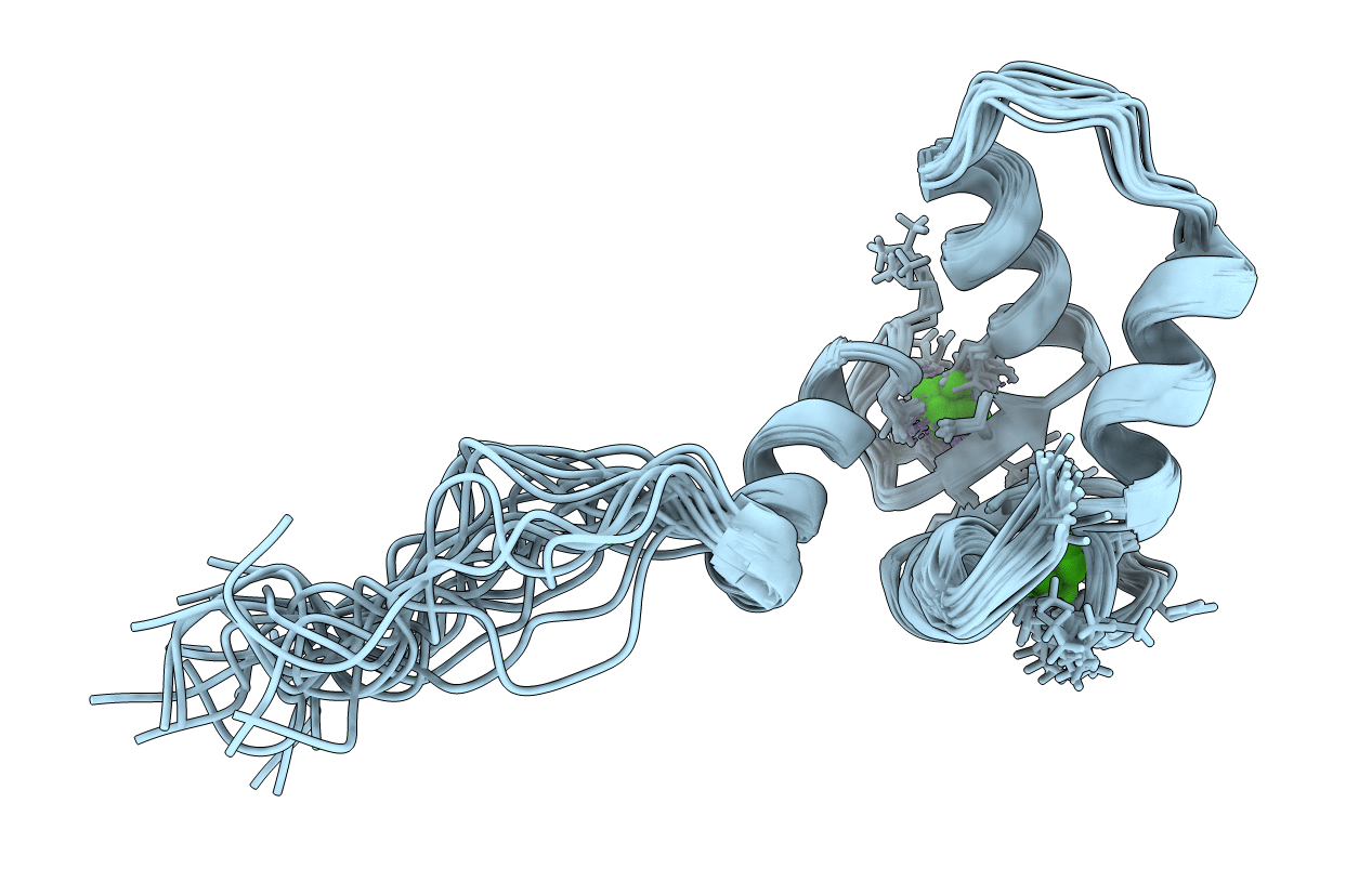
NMR STRUCTURE OF THE C TERMINAL DOMAIN OF CARDIAC TROPONIN C BOUND TO THE N TERMINAL DOMAIN OF CARDIAC TROPONIN I
Biological Source:
Source Organism(s):
Gallus gallus (Taxon ID: 9031)
Expression System(s):
Method Details:
Experimental Method:
Conformers Calculated:
20
Conformers Submitted:
20
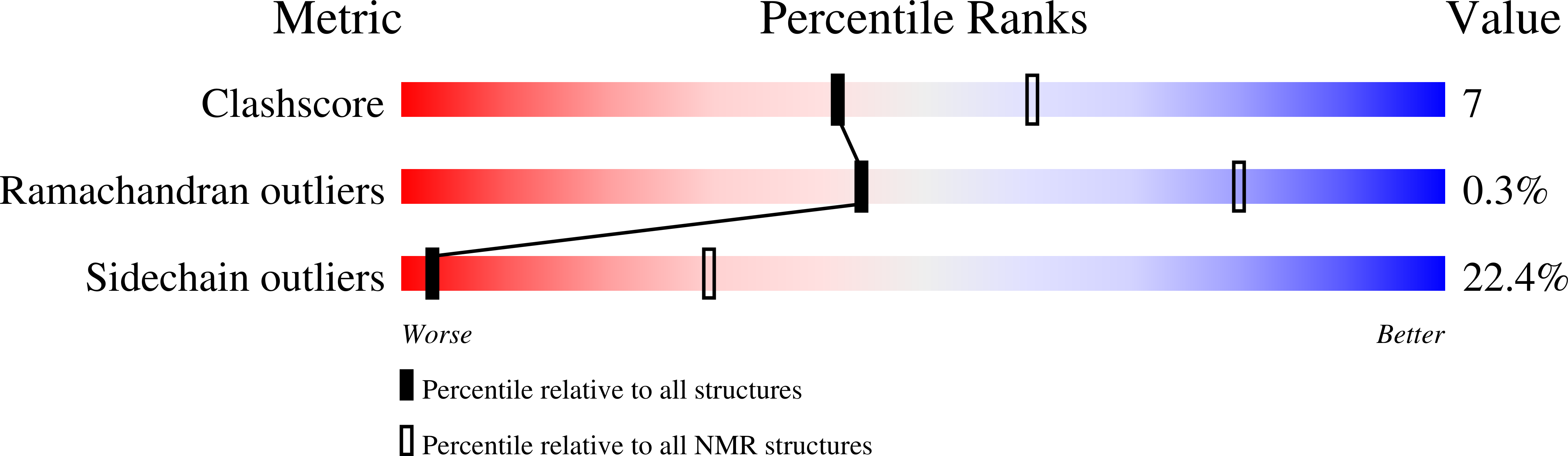
Selection Criteria:
structures with the lowest energy