Deposition Date
1994-01-06
Release Date
1994-04-30
Last Version Date
2024-02-14
Entry Detail
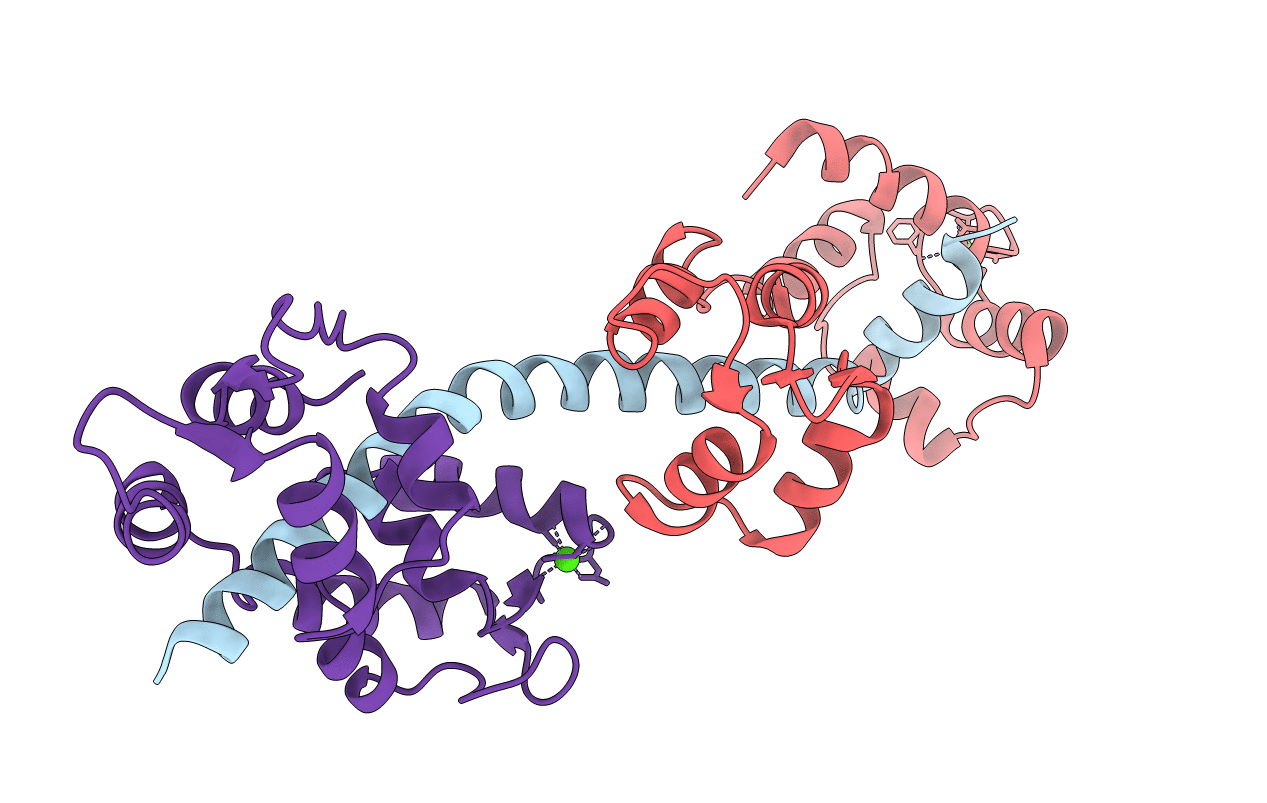
PDB ID:
1SCM
Keywords:
Title:
STRUCTURE OF THE REGULATORY DOMAIN OF SCALLOP MYOSIN AT 2.8 ANGSTROMS RESOLUTION
Biological Source:
Source Organism(s):
Argopecten irradians (Taxon ID: 31199)
Method Details:
Experimental Method:
Resolution:
2.80 Å
R-Value Work:
0.20
R-Value Observed:
0.20
Space Group:
P 1 21 1