Deposition Date
1993-08-23
Release Date
1994-01-31
Last Version Date
2024-02-14
Entry Detail
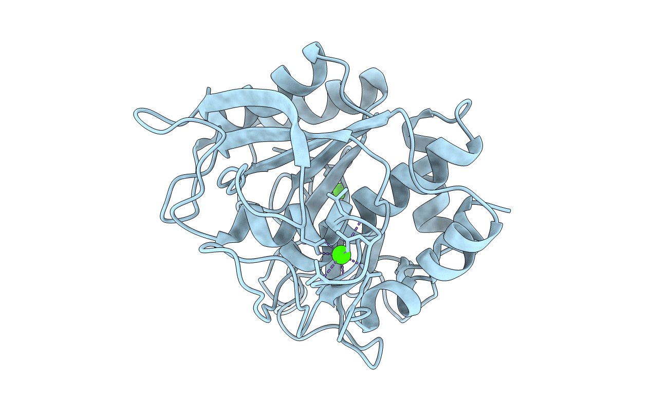
PDB ID:
1SCD
Keywords:
Title:
X-RAY CRYSTAL STRUCTURE OF CROSS-LINKED SUBTILISM CARLSBERG IN WATER VS. ACETONITRILE
Biological Source:
Source Organism(s):
Bacillus licheniformis (Taxon ID: 1402)
Method Details:
Experimental Method:
Resolution:
2.30 Å
R-Value Observed:
0.14
Space Group:
P 21 21 21