1SBW image
Entry Detail
PDB ID:
1SBW
Title:
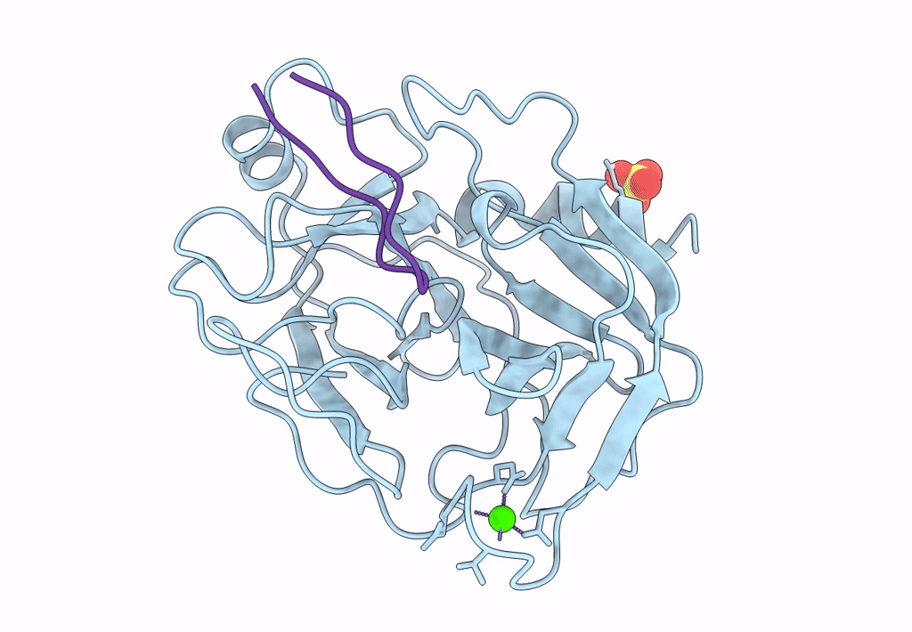
CRYSTAL STRUCTURE OF MUNG BEAN INHIBITOR LYSINE ACTIVE FRAGMENT COMPLEX WITH BOVINE BETA-TRYPSIN AT 1.8A RESOLUTION
Biological Source:
Source Organism:
PDB Version:
Deposition Date:
1999-04-29
Release Date:
1999-05-06
Method Details:
Experimental Method:
Resolution:
1.80 Å
R-Value Free:
0.19
R-Value Work:
0.16
Space Group:
P 21 21 21
Macromolecular Entities
Polymer Type:polypeptide(L)
Description:PROTEIN (BETA-TRYPSIN)
Chain IDs:A
Chain Length:223
Number of Molecules:1
Biological Source:Bos taurus
Polymer Type:polypeptide(L)
Description:PROTEIN (MUNG BEAN INHIBITOR LYSIN ACTIVE FRAGMENT)
Chain IDs:B (auth: I)
Chain Length:35
Number of Molecules:1
Biological Source:Vigna radiata
Primary Citation
Crystal structure of mung bean inhibitor lysine active fragment complex with bovine beta-trypsin at 1.8A resolution.
J.Biomol.Struct.Dyn. 16 1219 1224 (1999)
PMID: 10447205

Abstact

The crystal structure of the complex of mung bean inhibitor lysine active fragment with bovine beta-trypsin has been determined by X-ray crystallographic analysis at a resolution of 1.8 A. Refinement of the model of the complex converged at a final R value of 0.16. From the resulting electron density map, about one-third of the residues of the inhibitor were identified and two residues, at position P4 and P2' respectively, were found to be inconsistent with the sequence reported previously. The peptide chain of the inhibitor at the trypsin active site turns back sharply at Pro23I and forms a 9-residue reactive loop, which interacts with trypsin in a similar manner to the other families of inhibitors, suggesting an important and common role of these regions in exhibiting inhibitory activity.

Legend

Protein

Chemical

Disease

Primary Citation of related structures