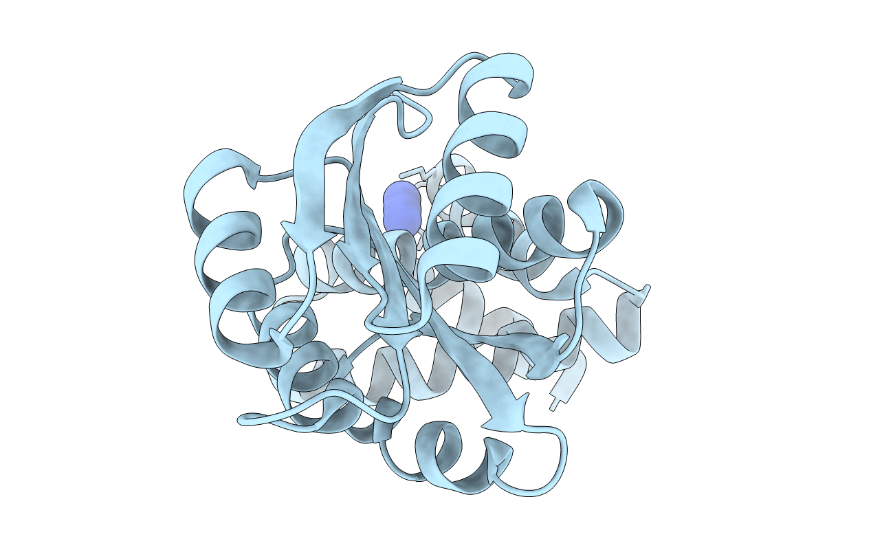
1S8N image
Entry Detail
PDB ID:
1S8N
Title:
Crystal structure of Rv1626 from Mycobacterium tuberculosis
Biological Source:
Host Organism:
PDB Version:
Deposition Date:
2004-02-03
Release Date:
2004-09-21
Method Details:
Experimental Method:
Resolution:
1.48 Å
R-Value Free:
0.22
R-Value Work:
0.20
R-Value Observed:
0.20
Space Group:
P 43 21 2
Macromolecular Entities
Polymer Type:polypeptide(L)
Description:putative antiterminator
Chain IDs:A
Chain Length:205
Number of Molecules:1
Biological Source:Mycobacterium tuberculosis
Ligand Molecules
Primary Citation
The Crystal and Solution Structure of a Putative Transcriptional Antiterminator from Mycobacterium tuberculosis.
Structure 12 1595 1605 (2004)
PMID: 15341725 DOI: 10.1016/j.str.2004.06.018

Abstact

We describe the crystal structure of Rv1626 from Mycobacterium tuberculosis at 1.48 A resolution and the corresponding solution structure determined from small angle X-ray scattering. The N-terminal domain shows structural homology to the receiver domains found in bacterial two-component systems. The C-terminal domain has high structural homology to a recently discovered RNA binding domain involved in transcriptional antitermination. The molecule in solution was found to be monomeric as it is in the crystal, but in solution it undergoes a conformational change that is triggered by changes in ionic strength. This is the first structure that links the phosphorylation cascade of the two-component systems with the antitermination event in the transcriptional machinery. Rv1626 belongs to a family of proteins, which we propose calling phosphorylation-dependent transcriptional antitermination regulators, so far only found in bacteria, and includes NasT, a protein from the assimilatory nitrate/nitrite reductase operon of Azetobacter vinelandii.

Legend

Protein

Chemical

Disease

Primary Citation of related structures