1S8G image
Deposition Date 2004-02-02
Release Date 2004-02-10
Last Version Date 2024-11-13
Entry Detail
PDB ID:
1S8G
Title:
Crystal structure of Lys49-Phospholipase A2 from Agkistrodon contortrix laticinctus, fatty acid bound form
Biological Source:
Source Organism:
Method Details:
Experimental Method:
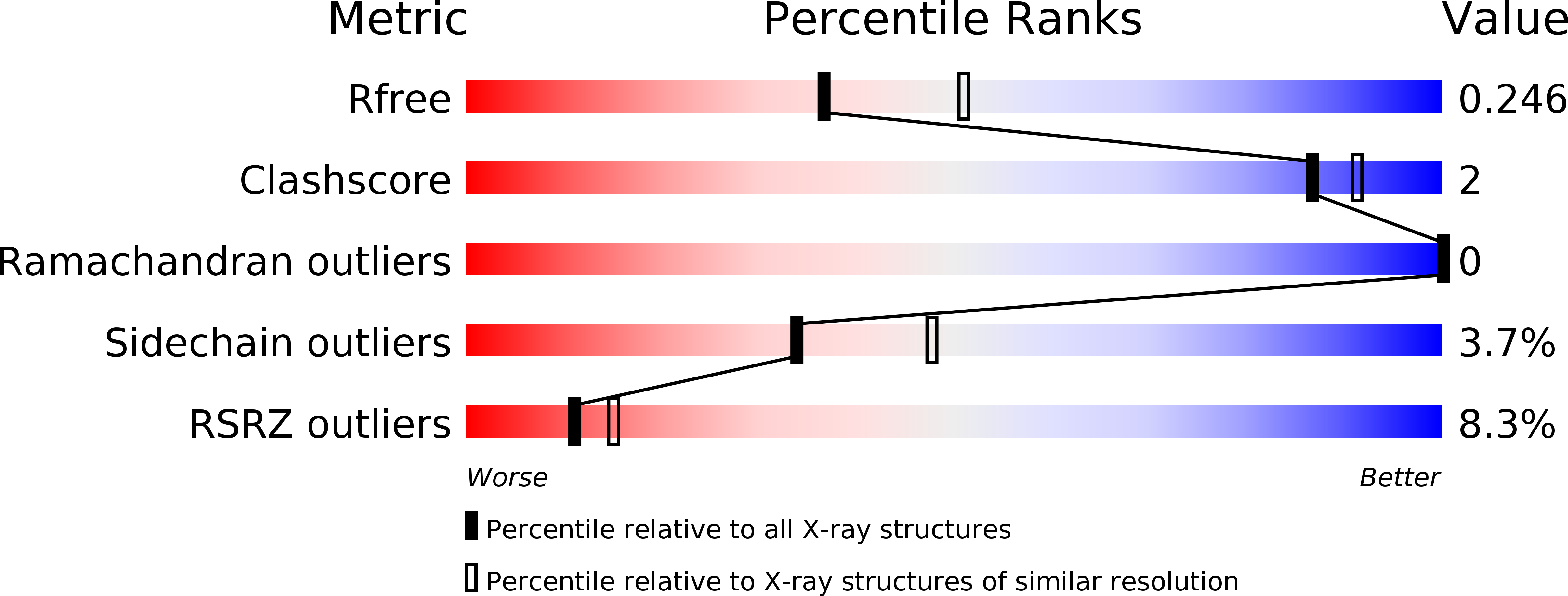
Resolution:
2.30 Å
R-Value Free:
0.24
R-Value Work:
0.20
R-Value Observed:
0.20
Space Group:
P 41 21 2
Macromolecular Entities
Structures with similar UniProt ID
Protein Blast
Polymer Type:polypeptide(L)
Molecule:Phospholipase A2 homolog
Chain IDs:A
Chain Length:121
Number of Molecules:1
Biological Source:Agkistrodon contortrix laticinctus
Primary Citation
A Molecular Mechanism for Lys49-Phospholipase A2 Activity Based on Ligand-induced Conformational Change.
J.Biol.Chem. 280 7326 7335 (2005)
PMID: 15596433 DOI: 10.1074/jbc.M410588200

Abstact

Agkistrodon contortrix laticinctus myotoxin is a Lys(49)-phospholipase A(2) (EC 3.1.1.4) isolated from the venom of the serpent A. contortrix laticinctus (broad-banded copperhead). We present here three monomeric crystal structures of the myotoxin, obtained under different crystallization conditions. The three forms present notable structural differences and reveal that the presence of a ligand in the active site (naturally presumed to be a fatty acid) induces the exposure of a hydrophobic surface (the hydrophobic knuckle) toward the C terminus. The knuckle in A. contortrix laticinctus myotoxin involves the side chains of Phe(121) and Phe(124) and is a consequence of the formation of a canonical structure for the main chain within the region of residues 118-125. Comparison with other Lys(49)-phospholipase A(2) myotoxins shows that although the knuckle is a generic structural motif common to all members of the family, it is not readily recognizable by simple sequence analyses. An activation mechanism is proposed that relates fatty acid retention at the active site to conformational changes within the C-terminal region, a part of the molecule that has long been associated with Ca(2+)-independent membrane damaging activity and myotoxicity. This provides, for the first time, a direct structural connection between the phospholipase "active site" and the C-terminal "myotoxic site," justifying the otherwise enigmatic conservation of the residues of the former in supposedly catalytically inactive molecules.

Legend

Protein

Chemical

Disease

Primary Citation of related structures
Feedback Form
Name
Email
Institute
Feedback