Deposition Date
2004-02-02
Release Date
2004-06-11
Last Version Date
2023-08-23
Entry Detail
PDB ID:
1S8F
Keywords:
Title:
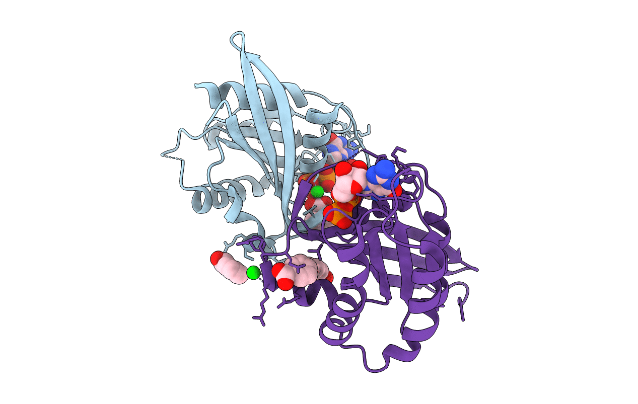
Crystal structure of Rab9 complexed to GDP reveals a dimer with an active conformation of switch II
Biological Source:
Source Organism(s):
Canis lupus familiaris (Taxon ID: 9615)
Expression System(s):
Method Details:
Experimental Method:
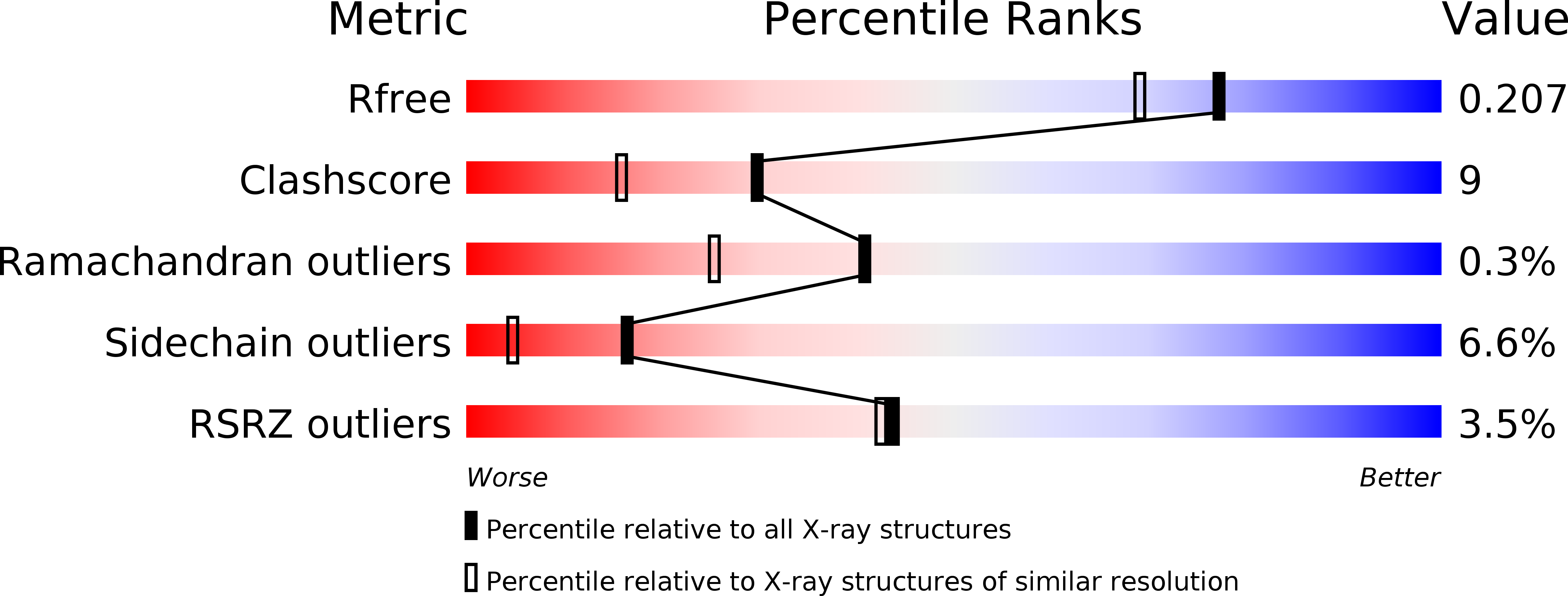
Resolution:
1.77 Å
R-Value Free:
0.22
R-Value Work:
0.16
Space Group:
I 41