Deposition Date
2004-02-02
Release Date
2004-08-03
Last Version Date
2023-08-23
Entry Detail
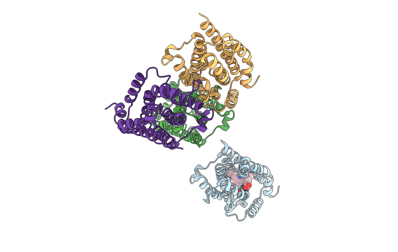
PDB ID:
1S8C
Keywords:
Title:
Crystal structure of human heme oxygenase in a complex with biliverdine
Biological Source:
Source Organism(s):
Homo sapiens (Taxon ID: 9606)
Expression System(s):
Method Details:
Experimental Method:
Resolution:
2.19 Å
R-Value Free:
0.28
R-Value Work:
0.24
Space Group:
P 1 21 1