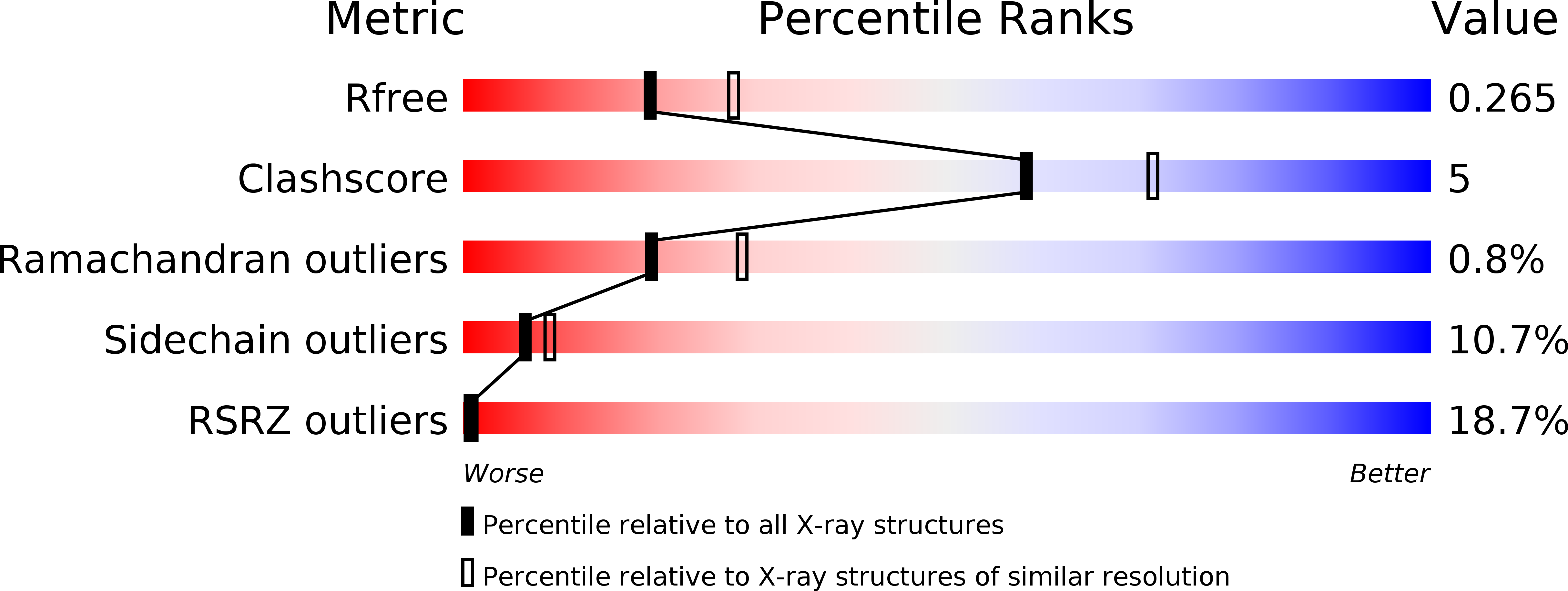
1S5J image
Deposition Date 2004-01-21
Release Date 2004-11-09
Last Version Date 2024-10-30
Entry Detail
PDB ID:
1S5J
Keywords:
Title:
Insight in DNA Replication: The crystal structure of DNA Polymerase B1 from the archaeon Sulfolobus solfataricus
Biological Source:
Source Organism:
Method Details:
Experimental Method:
Resolution:
2.40 Å
R-Value Free:
0.27
R-Value Work:
0.23
R-Value Observed:
0.23
Space Group:
C 1 2 1
Macromolecular Entities
Polymer Type:polypeptide(L)
Molecule:DNA polymerase I
Gene (Uniprot):dpo1
Chain IDs:A
Chain Length:847
Number of Molecules:1
Biological Source:Sulfolobus solfataricus
Primary Citation
Insights into DNA replication: the crystal structure of DNA polymerase B1 from the archaeon Sulfolobus solfataricus
Structure 12 2001 2008 (2004)
PMID: 15530364 DOI: 10.1016/j.str.2004.09.007

Abstact

To minimize the large number of mispairs during genome duplication owing to the large amount of DNA to be synthesized, many replicative polymerases have accessory domains with complementary functions. We describe the crystal structure of replicative DNA polymerase B1 from the archaeon Sulfolobus solfataricus. Comparison between other known structures indicates that although the protein is folded into the typical N-terminal, editing 3'-5'exonuclease, and C-terminal right-handed polymerase domains, it is characterized by the unusual presence of two extra alpha helices in the N-terminal domain interacting with the fingers helices to form an extended fingers subdomain, a structural feature that can account for some functional features of the protein. We explore the structural basis of specific lesion recognition, the initial step in DNA repair, describing how the N-terminal subdomain pocket of archaeal DNA polymerases could allow specific recognition of deaminated bases such as uracil and hypoxanthine in addition to the typical DNA bases.

Legend

Protein

Chemical

Disease

Primary Citation of related structures