1S5H image
Entry Detail
PDB ID:
1S5H
Title:
Potassium Channel Kcsa-Fab Complex T75C mutant in K+
Biological Source:
PDB Version:
Deposition Date:
2004-01-20
Release Date:
2004-05-18
Method Details:
Experimental Method:
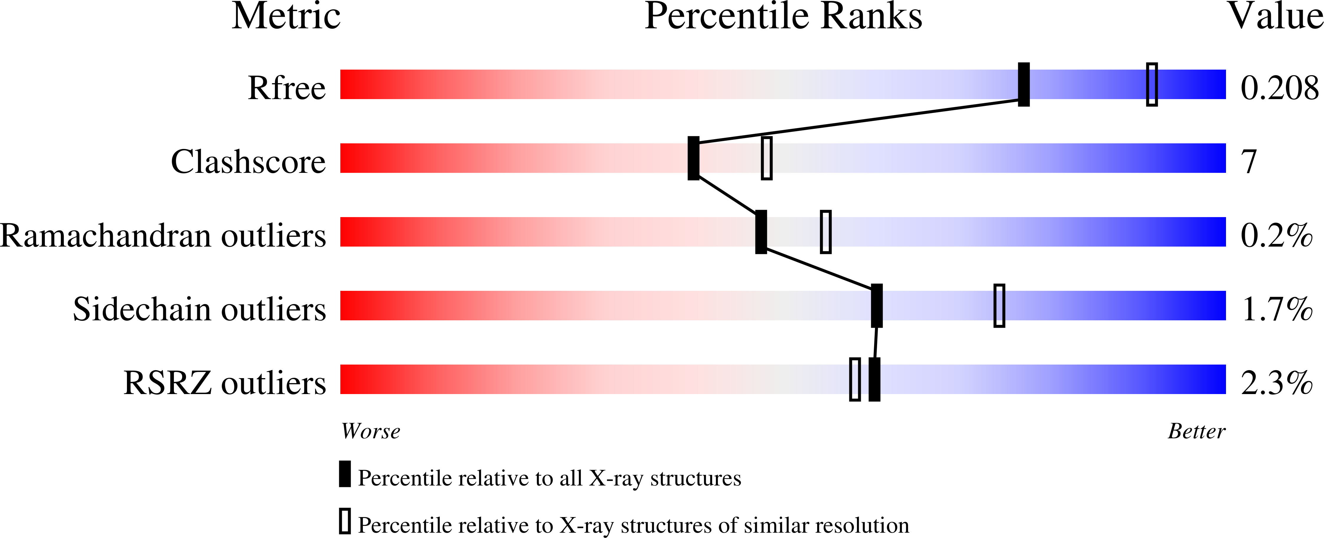
Resolution:
2.20 Å
R-Value Free:
0.24
R-Value Work:
0.22
R-Value Observed:
0.22
Space Group:
I 4
Macromolecular Entities
Polymer Type:polypeptide(L)
Description:ANTIBODY FAB FRAGMENT LIGHT CHAIN
Chain IDs:A
Chain Length:212
Number of Molecules:1
Biological Source:Mus musculus
Polymer Type:polypeptide(L)
Description:ANTIBODY FAB FRAGMENT HEAVY CHAIN
Chain IDs:B
Chain Length:219
Number of Molecules:1
Biological Source:Mus musculus
Polymer Type:polypeptide(L)
Description:Voltage-gated potassium channel
Mutations:P2A, T75C
Chain IDs:C
Chain Length:124
Number of Molecules:1
Biological Source:Streptomyces coelicolor, Streptomyces lividans
Primary Citation
A mutant KcsA K(+) channel with altered conduction properties and selectivity filter ion distribution.
J.Mol.Biol. 338 839 846 (2004)
PMID: 15099749 DOI: 10.1016/j.jmb.2004.03.020

Abstact

The selectivity filter of K(+) channels is comprised of a linear queue of four equal-spaced ion-binding sites spanning a distance of 12A. Each site is formed of eight oxygen atoms from the protein. The first three sites, numbered 1-3 from the extracellular side, are made of exclusively main-chain carbonyl oxygen atoms. The fourth site, closest to the intracellular side, is made of four main-chain carbonyl oxygen atoms and four threonine side-chain hydroxyl oxygen atoms. Here we characterize the effects of mutating the threonine to cysteine on the distribution of ions in the selectivity filter and on the conduction of ions through the filter. The mutation influences the occupancy of K(+) at sites 2 and 4 and it reduces the maximum rate of conduction in the limit of high K(+) concentration. The mutation does not affect the conduction of Rb(+). These results can be understood in the context of a conduction mechanism in which a pair of K ions switch between energetically balanced 1,3 and 2,4 configurations.

Legend

Protein

Chemical

Disease

Primary Citation of related structures