1S4N image
Deposition Date 2004-01-16
Release Date 2004-05-04
Last Version Date 2024-10-16
Entry Detail
PDB ID:
1S4N
Keywords:
Title:
Crystal structure of yeast alpha1,2-mannosyltransferase Kre2p/Mnt1p
Biological Source:
Source Organism:
Host Organism:
Method Details:
Experimental Method:
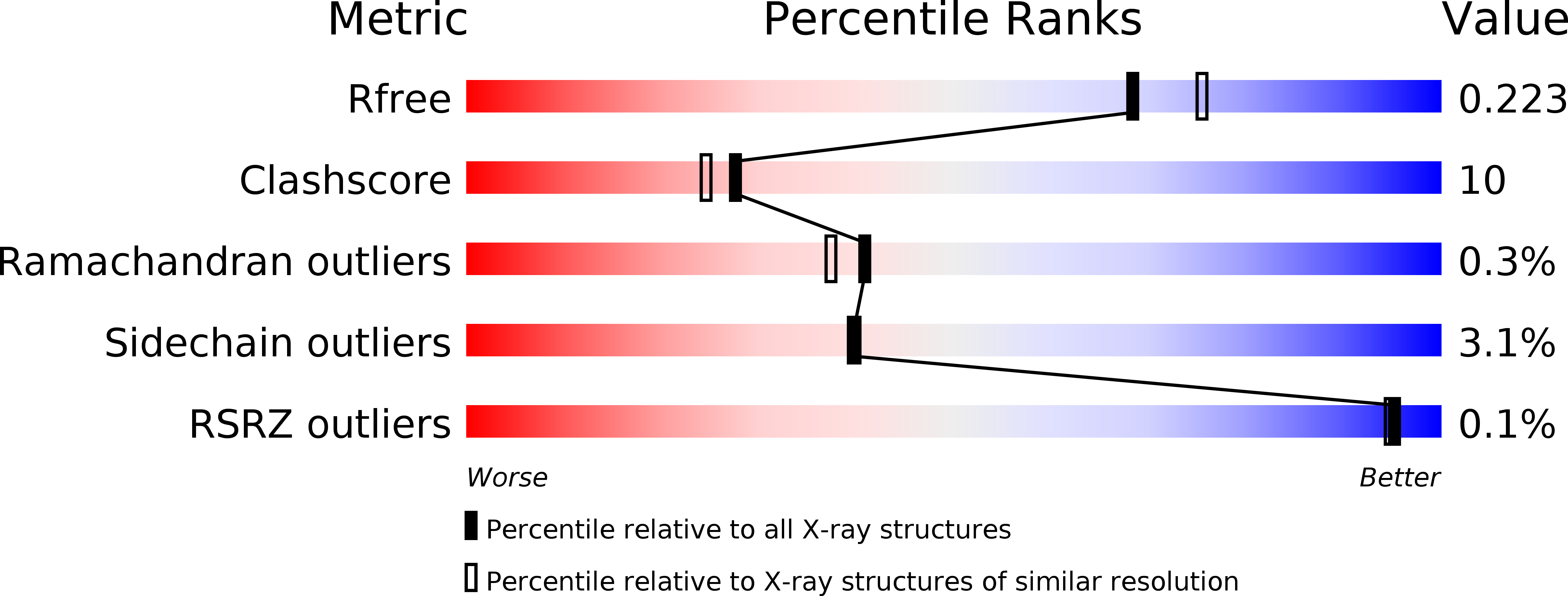
Resolution:
2.01 Å
R-Value Free:
0.23
R-Value Work:
0.18
R-Value Observed:
0.18
Space Group:
P 1 21 1
Macromolecular Entities
Polymer Type:polypeptide(L)
Molecule:Glycolipid 2-alpha-mannosyltransferase
Gene (Uniprot):KRE2
Chain IDs:A, B
Chain Length:348
Number of Molecules:2
Biological Source:Saccharomyces cerevisiae
Modified Residue
Compound ID Chain ID Parent Comp ID Details 2D Image
ASN A ASN GLYCOSYLATION SITE
Primary Citation
Structure of Kre2p/Mnt1p: A YEAST {alpha}1,2-MANNOSYLTRANSFERASE INVOLVED IN MANNOPROTEIN BIOSYNTHESIS
J.Biol.Chem. 279 17921 17931 (2004)
PMID: 14752117 DOI: 10.1074/jbc.M312720200

Abstact

Kre2p/Mnt1p is a Golgi alpha1,2-mannosyltransferase involved in the biosynthesis of Saccharomyces cerevisiae cell wall glycoproteins. The protein belongs to glycosyltransferase family 15, a member of which has been implicated in virulence of Candida albicans. We present the 2.0 A crystal structures of the catalytic domain of Kre2p/Mnt1p and its binary and ternary complexes with GDP/Mn(2+) and GDP/Mn(2+)/acceptor methyl-alpha-mannoside. The protein has a mixed alpha/beta fold similar to the glycosyltransferase-A (GT-A) fold. Although the GDP-mannose donor was used in the crystallization experiments and the GDP moiety is bound tightly to the active site, the mannose is not visible in the electron density. The manganese is coordinated by a modified DXD motif (EPD), with only the first glutamate involved in a direct interaction. The position of the donor mannose was modeled using the binary and ternary complexes of other GT-A enzymes. The C1" of the modeled donor mannose is within hydrogen-bonding distance of both the hydroxyl of Tyr(220) and the O2 of the acceptor mannose. The O2 of the acceptor mannose is also within hydrogen bond distance of the hydroxyl of Tyr(220). The structures, modeling, site-directed mutagenesis, and kinetic analysis suggest two possible catalytic mechanisms. Either a double-displacement mechanism with the hydroxyl of Tyr(220) as the potential nucleophile or alternatively, an S(N)i-like mechanism with Tyr(220) positioning the substrates for catalysis. The importance of Tyr(220) in both mechanisms is highlighted by a 3000-fold reduction in k(cat) in the Y220F mutant.

Legend

Protein

Chemical

Disease

Primary Citation of related structures