
Deposition Date
2004-01-13
Release Date
2004-09-07
Last Version Date
2023-08-23
Entry Detail
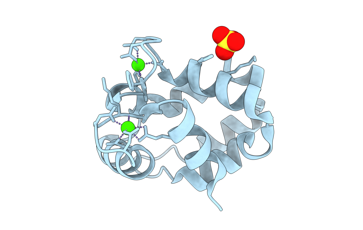
PDB ID:
1S3P
Keywords:
Title:
Crystal structure of rat alpha-parvalbumin S55D/E59D mutant
Biological Source:
Source Organism(s):
Rattus norvegicus (Taxon ID: 10116)
Expression System(s):
Method Details:
Experimental Method:
Resolution:
2.00 Å
R-Value Free:
0.22
R-Value Work:
0.18
R-Value Observed:
0.18
Space Group:
P 21 21 2
