
Deposition Date
2004-01-13
Release Date
2004-01-27
Last Version Date
2021-10-27
Entry Detail
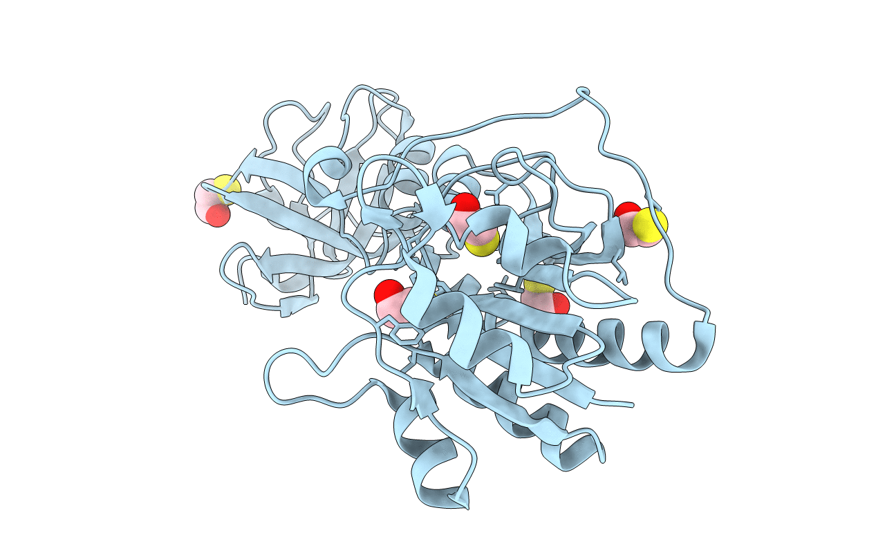
PDB ID:
1S3I
Keywords:
Title:
Crystal structure of the N terminal hydrolase domain of 10-formyltetrahydrofolate dehydrogenase
Biological Source:
Source Organism(s):
Rattus norvegicus (Taxon ID: 10116)
Expression System(s):
Method Details:
Experimental Method:
Resolution:
2.30 Å
R-Value Free:
0.30
R-Value Work:
0.24
R-Value Observed:
0.24
Space Group:
P 21 21 2
