Deposition Date
2004-01-12
Release Date
2004-04-20
Last Version Date
2024-02-14
Entry Detail
PDB ID:
1S35
Keywords:
Title:
Crystal Structure of Repeats 8 and 9 of Human Erythroid Spectrin
Biological Source:
Source Organism(s):
Homo sapiens (Taxon ID: 9606)
Expression System(s):
Method Details:
Experimental Method:
Resolution:
2.40 Å
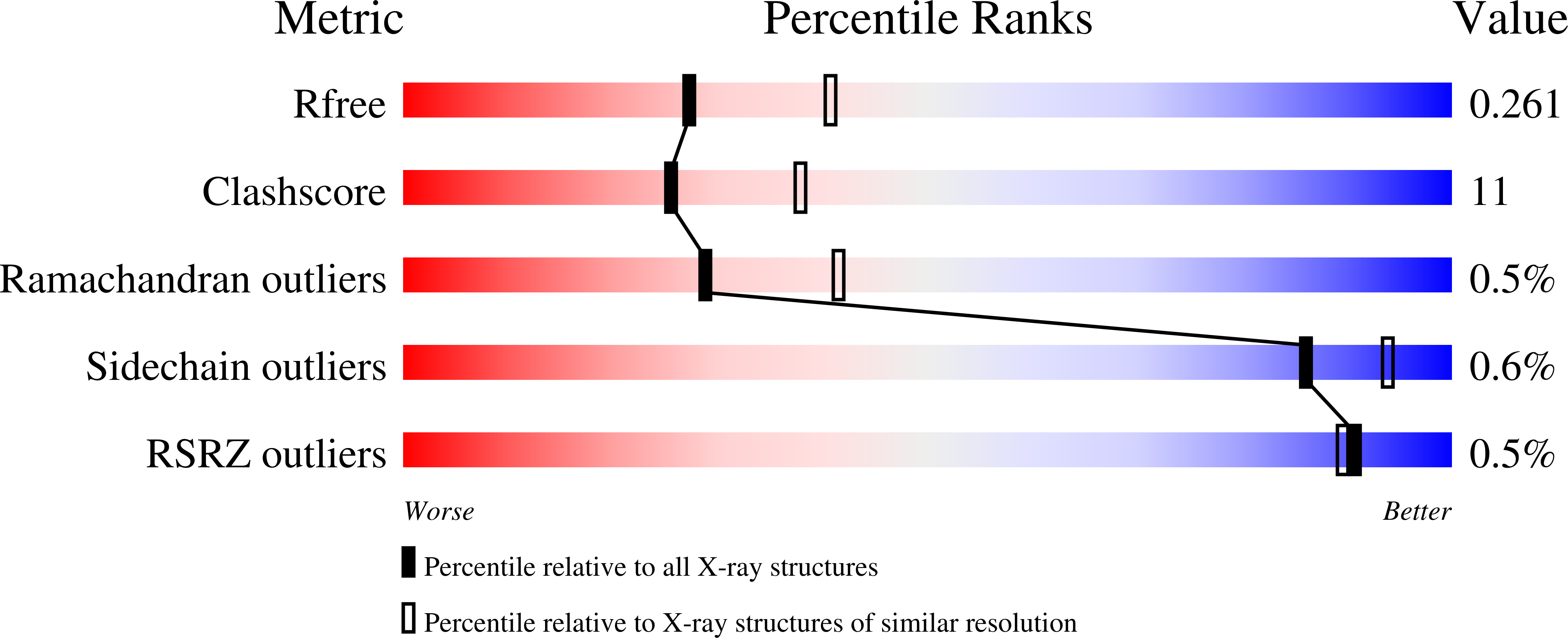
R-Value Free:
0.25
R-Value Work:
0.22
Space Group:
P 41 21 2