1S30 image
Deposition Date 2004-01-12
Release Date 2004-06-22
Last Version Date 2023-08-23
Entry Detail
PDB ID:
1S30
Title:
X-ray crystal structure of Desulfovibrio vulgaris Rubrerythrin with displacement of iron by zinc at the diiron Site
Biological Source:
Source Organism:
Method Details:
Experimental Method:
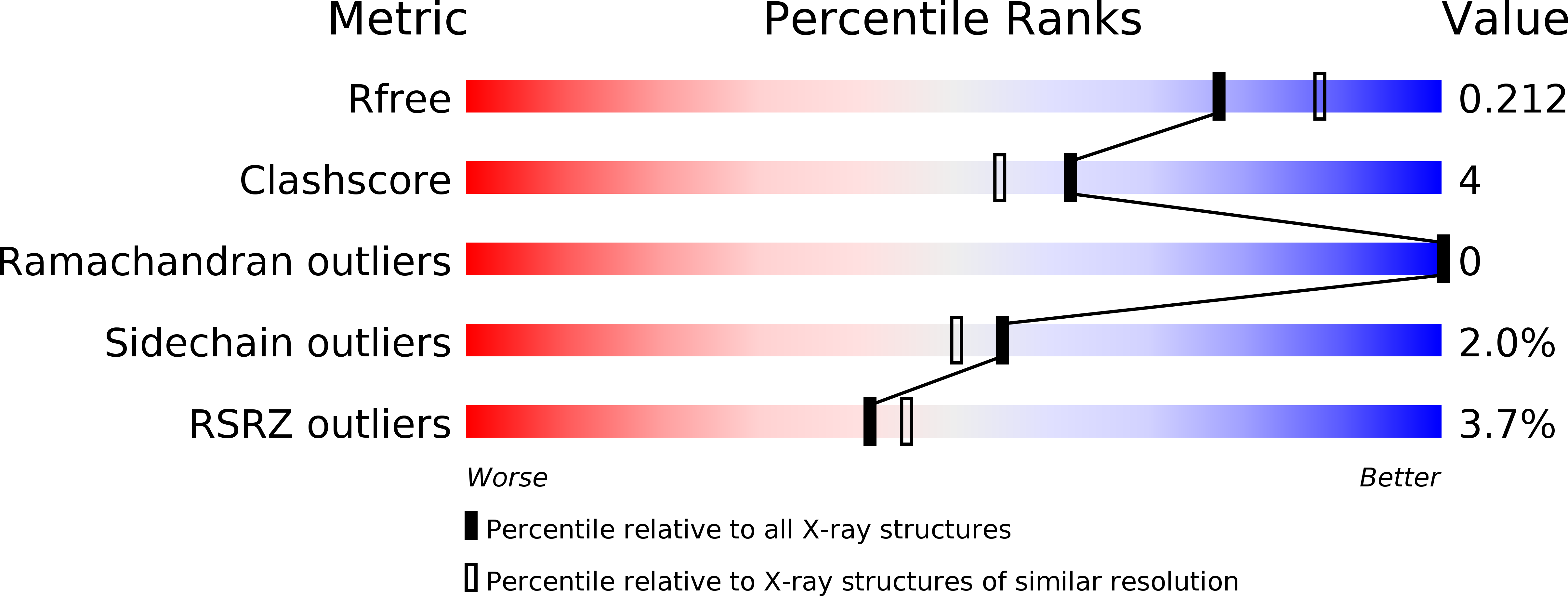
Resolution:
2.05 Å
R-Value Free:
0.20
R-Value Work:
0.17
R-Value Observed:
0.17
Space Group:
I 2 2 2
Macromolecular Entities
Structures with similar UniProt ID
Protein Blast
Polymer Type:polypeptide(L)
Molecule:Rubrerythrin
Gene (Uniprot):rbr
Chain IDs:A
Chain Length:191
Number of Molecules:1
Biological Source:Desulfovibrio vulgaris
Primary Citation
Displacement of iron by zinc at the diiron site of Desulfovibrio vulgaris rubrerythrin: X-ray crystal structure and anomalous scattering analysis
J.Inorg.Biochem. 98 786 796 (2004)
PMID: 15134924 DOI: 10.1016/j.jinorgbio.2004.01.005

Abstact

X-ray crystal structures of recombinant Desulfovibrio (D.) vulgaris rubrerythrin (Rbr) have shown a diiron site, whereas the crystal structure of Rbr "as-isolated" from D. vulgaris was reported to contain a mixed Zn,Fe binuclear site. To investigate the possibility that zinc had displaced iron during isolation or crystallization of the "as-isolated" D. vulgaris Rbr, the X-ray crystal structure of recombinant D. vulgaris all-iron Rbr that had been incubated with excess zinc sulfate prior to crystallization, yielding a protein labeled Zn,FeRbr, was solved. Analysis of the anomalous scattering data obtained at two different wavelengths showed that zinc had displaced a significant proportion of iron from both iron centers of the diiron site, and that no iron had been displaced from the [Fe(SCys)(4)] site. UV-visible absorption spectra of the redissolved Zn,FeRbr crystals showed 30-40% retention of oxo-bridged diferric sites, and the redissolved crystals had 37% of the peroxidase specific activity of the starting all-iron Rbr, which, together with the crystallographic results, indicate a predominant mixture of Fe1,Fe2 and Zn1,Zn2 sites. The structure of the Zn(Fe)1,Fe(Zn)2 binuclear site in the Zn,FeRbr crystals was very similar to that of the Zn,Fe binuclear site reported for the "as-isolated" D. vulgaris Rbr, including tetrahedral four-coordination at the Zn(Fe)1 site. The diiron sites in the recombinant Zn,FeRbr crystals were likely at least partially reduced during synchrotron irradiation. Our results suggest that the mixed-metal binuclear site reported for the "as-isolated" D. vulgaris Rbr could be due to displacement of iron from a native diiron site by adventitious zinc during isolation and/or crystallization, and that reduced diiron and dizinc sites can adopt very similar structures in Rbr.

Legend

Protein

Chemical

Disease

Primary Citation of related structures
Feedback Form
Name
Email
Institute
Feedback