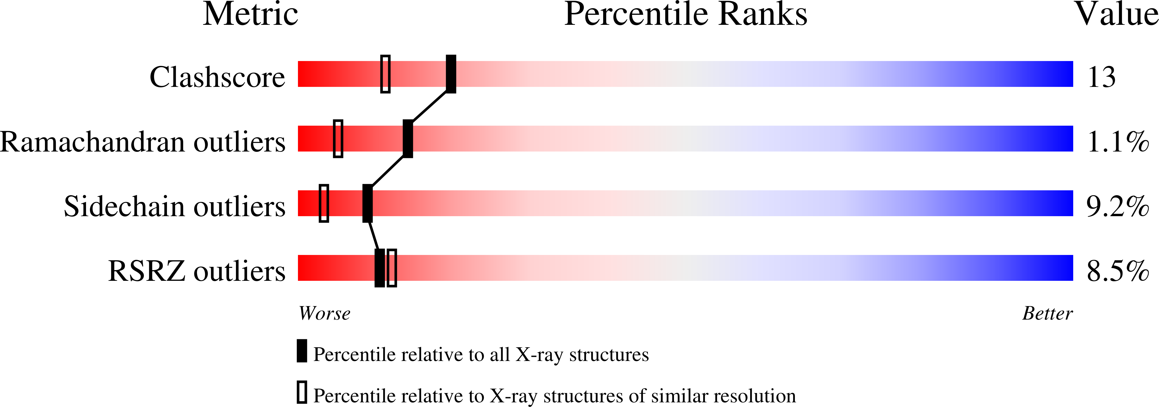
1S2X image
Deposition Date 2004-01-12
Release Date 2004-07-27
Last Version Date 2024-11-20
Entry Detail
PDB ID:
1S2X
Title:
Crystal structure of Cag-Z from Helicobacter pylori
Biological Source:
Source Organism:
Method Details:
Experimental Method:
Resolution:
1.90 Å
R-Value Free:
0.26
R-Value Work:
0.23
R-Value Observed:
0.22
Space Group:
P 21 21 21
Macromolecular Entities
Structures with similar UniProt ID
Protein Blast
Polymer Type:polypeptide(L)
Molecule:Cag-Z
Gene (Uniprot):cag-Z
Mutagens:M1D
Chain IDs:A
Chain Length:206
Number of Molecules:1
Biological Source:Helicobacter pylori
Modified Residue
Compound ID Chain ID Parent Comp ID Details 2D Image
MSE A MET SELENOMETHIONINE
Ligand Molecules
Primary Citation
Crystal structure of CagZ, a protein from the Helicobacter pylori pathogenicity island that encodes for a type IV secretion system
J.Mol.Biol. 340 881 889 (2004)
PMID: 15223328 DOI: 10.1016/j.jmb.2004.05.016

Abstact

CagZ, a 23 kDa protein encoded by the cagZ gene (HP0526) of the cag pathogenicity island of Helicobacter pylori, has been cloned, over-expressed, purified and its three-dimensional structure determined. The protein consists of a single compact L-shaped domain, composed of seven alpha-helices including about 70% of the total residues. Three-dimensional homology searches did not reveal structural homologues, and CagZ can be considered representative of a new protein fold. The presence of a disordered C-terminal tail and the nature of the molecular surface suggest that CagZ may participate in the interaction of effector proteins with one or more components of the H.pylori type IV secretion system on the cytoplasmic side of the inner membrane.

Legend

Protein

Chemical

Disease

Primary Citation of related structures