Deposition Date
2004-01-07
Release Date
2004-06-15
Last Version Date
2021-10-27
Entry Detail
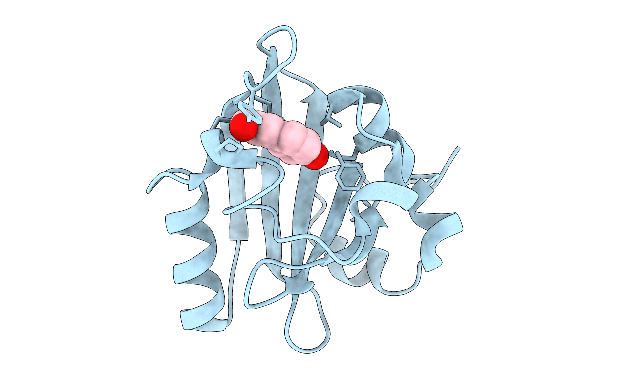
PDB ID:
1S1Z
Keywords:
Title:
Photoactivated chromophore conformation in Photoactive Yellow Protein (E46Q mutant) from 10 to 500 nanoseconds
Biological Source:
Source Organism(s):
Halorhodospira halophila (Taxon ID: 1053)
Expression System(s):
Method Details:
Experimental Method:
Resolution:
1.60 Å
R-Value Free:
0.06
R-Value Work:
0.05
R-Value Observed:
0.05
Space Group:
P 63