Deposition Date
2004-01-06
Release Date
2004-05-04
Last Version Date
2024-02-14
Entry Detail
PDB ID:
1S1J
Keywords:
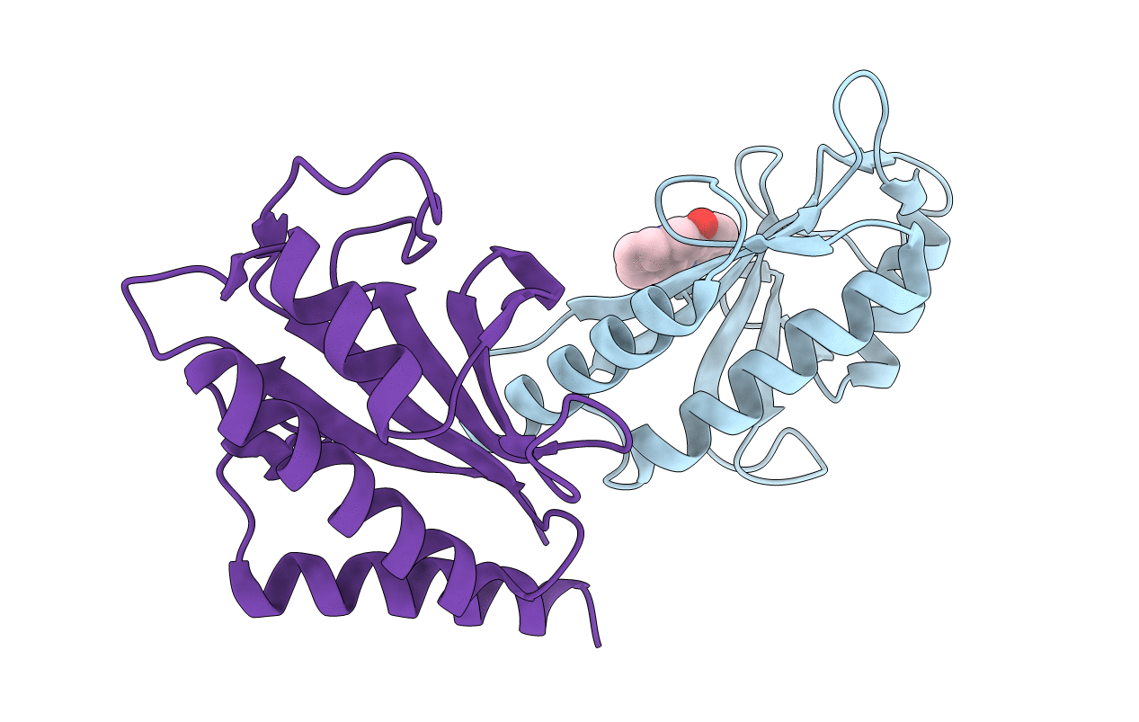
Title:
Crystal Structure of ZipA in complex with indoloquinolizin inhibitor 1
Biological Source:
Source Organism:
Escherichia coli (Taxon ID: 562)
Host Organism:
Method Details:
Experimental Method:
Resolution:
2.18 Å
R-Value Free:
0.23
R-Value Work:
0.19
R-Value Observed:
0.19
Space Group:
P 1 21 1