
Deposition Date
2003-12-30
Release Date
2004-03-16
Last Version Date
2023-08-23
Entry Detail
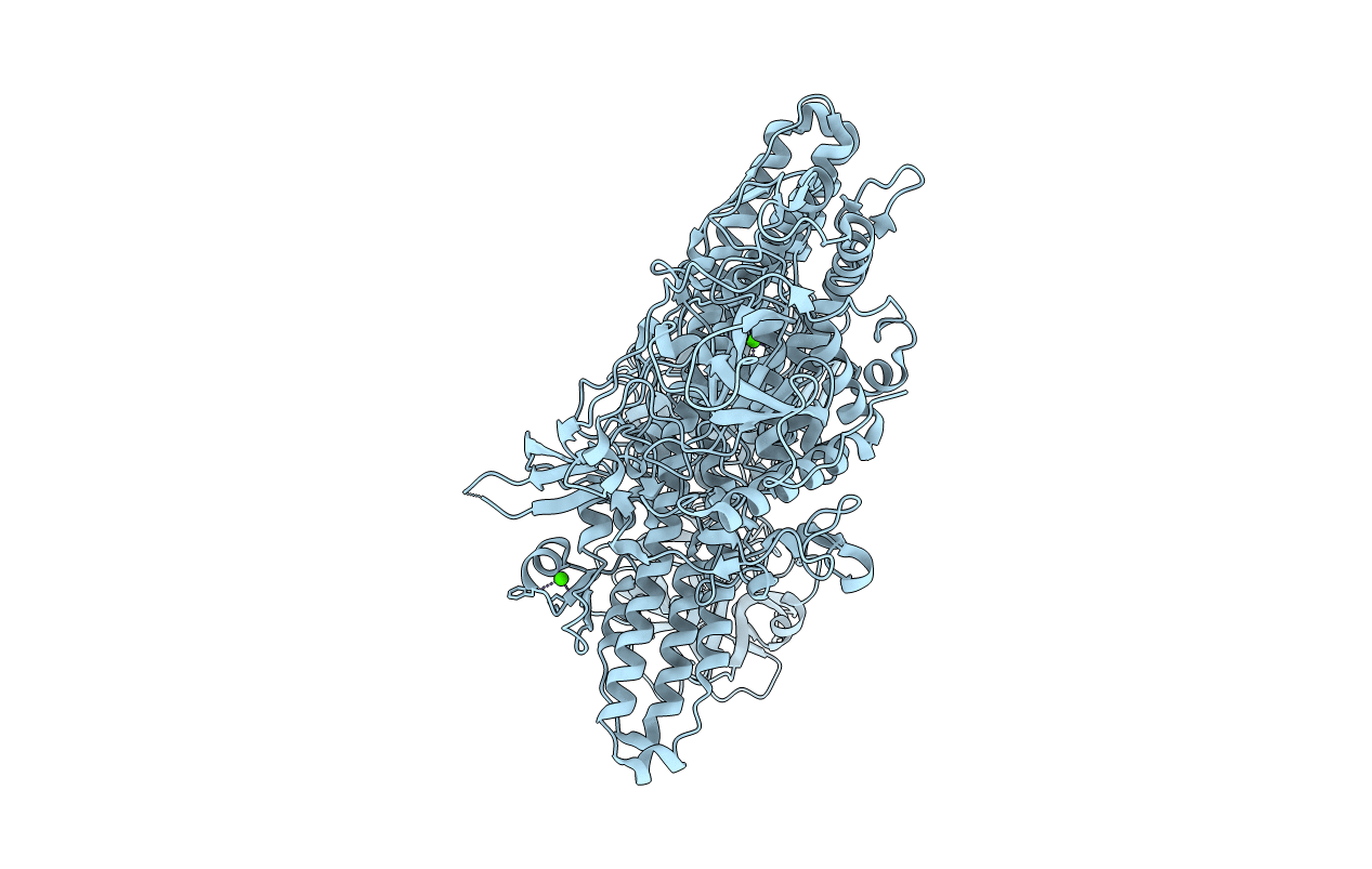
PDB ID:
1S0B
Keywords:
Title:
Crystal structure of botulinum neurotoxin type B at pH 4.0
Biological Source:
Source Organism(s):
Clostridium botulinum (Taxon ID: 1491)
Method Details:
Experimental Method:
Resolution:
2.00 Å
R-Value Free:
0.23
R-Value Work:
0.20
R-Value Observed:
0.20
Space Group:
P 1 21 1
