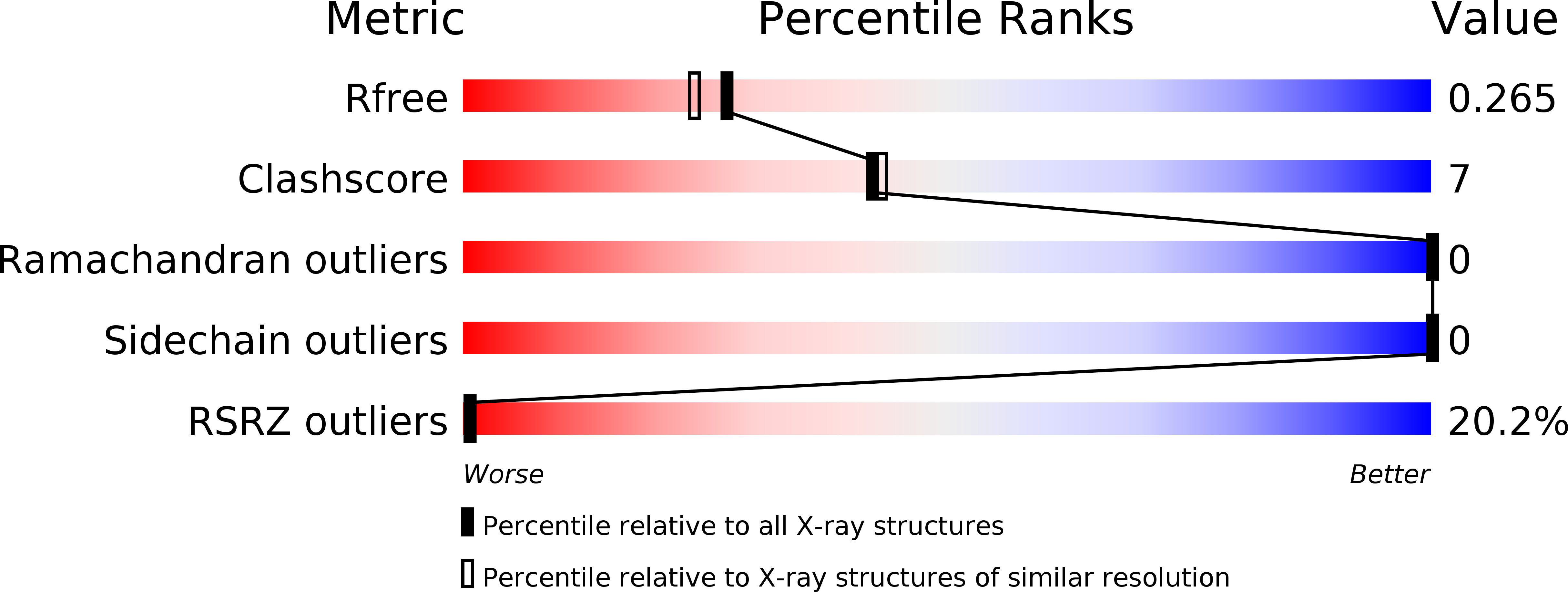
1RZX image
Deposition Date 2003-12-29
Release Date 2004-01-27
Last Version Date 2024-10-30
Entry Detail
PDB ID:
1RZX
Keywords:
Title:
Crystal Structure of a Par-6 PDZ-peptide Complex
Biological Source:
Source Organism:
Method Details:
Experimental Method:
Resolution:
2.10 Å
R-Value Free:
0.27
R-Value Work:
0.22
R-Value Observed:
0.22
Space Group:
P 63
Macromolecular Entities
Structures with similar UniProt ID
Protein Blast
Polymer Type:polypeptide(L)
Molecule:CG5884-PA
Gene (Uniprot):par-6
Chain IDs:A
Chain Length:98
Number of Molecules:1
Biological Source:Drosophila melanogaster
Polymer Type:polypeptide(L)
Molecule:Acetylated VKESLV Peptide
Chain IDs:B
Chain Length:7
Number of Molecules:1
Biological Source:
Primary Citation
Cdc42 regulates the Par-6 PDZ domain through an allosteric CRIB-PDZ transition.
Mol.Cell 13 665 676 (2004)
PMID: 15023337 DOI: 10.1016/S1097-2765(04)00086-3

Abstact

Regulation of protein interaction domains is required for cellular signaling dynamics. Here, we show that the PDZ protein interaction domain from the cell polarity protein Par-6 is regulated by the Rho GTPase Cdc42. Cdc42 binds to a CRIB domain adjacent to the PDZ domain, increasing the affinity of the Par-6 PDZ for its carboxy-terminal ligand by approximately 13-fold. Par-6 PDZ regulation is required for function as mutational disruption of Cdc42-Par-6 PDZ coupling leads to inactivation of Par-6 in polarized MDCK epithelial cells. Structural analysis reveals that the free PDZ domain has several deviations from the canonical PDZ conformation that account for its low ligand affinity. Regulation results from a Cdc42-induced conformational transition in the CRIB-PDZ module that causes the PDZ to assume a canonical, high-affinity PDZ conformation. The coupled CRIB and PDZ architecture of Par-6 reveals how simple binding domains can be combined to yield complex regulation.

Legend

Protein

Chemical

Disease

Primary Citation of related structures