1RYX image
Deposition Date 2003-12-23
Release Date 2004-07-13
Last Version Date 2024-11-20
Entry Detail
PDB ID:
1RYX
Keywords:
Title:
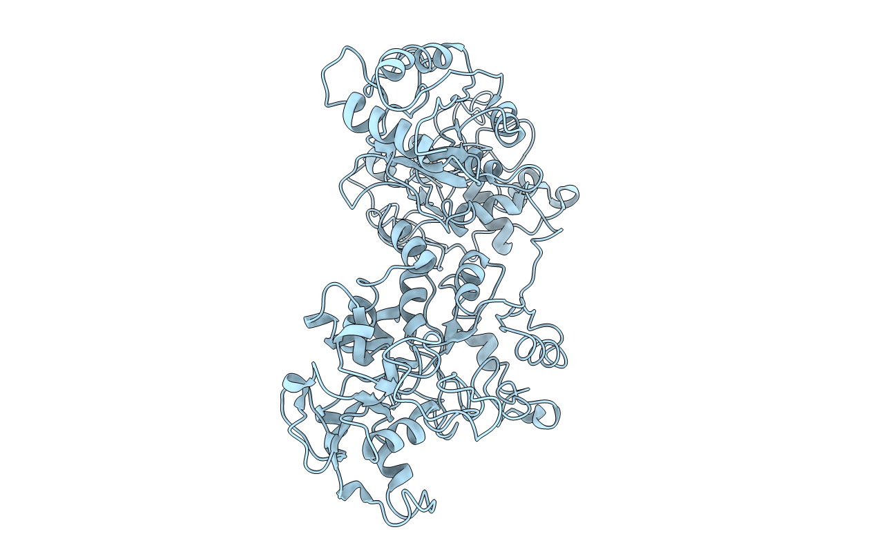
Crystal structure of hen serum transferrin in apo- form
Biological Source:
Source Organism:
Gallus gallus (Taxon ID: 9031)
Method Details:
Experimental Method:
Resolution:
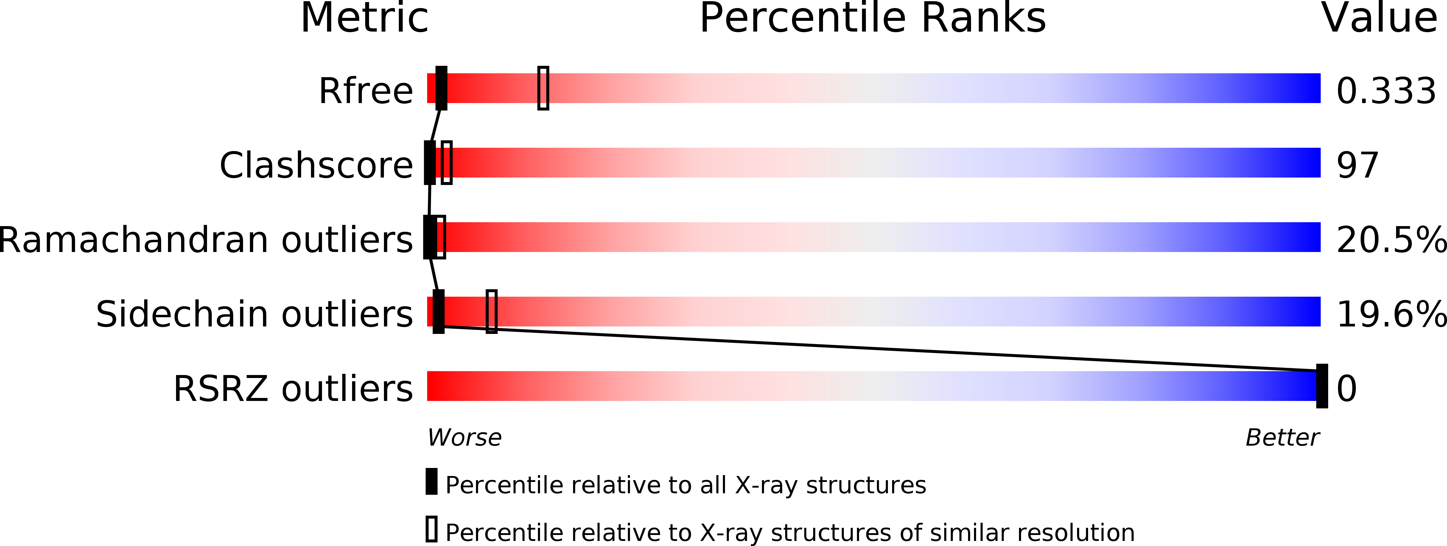
3.50 Å
R-Value Free:
0.34
R-Value Work:
0.28
R-Value Observed:
0.28
Space Group:
P 43 21 2
Macromolecular Entities
Polymer Type:polypeptide(L)
Molecule:Ovotransferrin
Chain IDs:A
Chain Length:686
Number of Molecules:1
Biological Source:Gallus gallus
Ligand Molecules
Primary Citation
Tertiary structural changes associated with iron binding and release in hen serum transferrin: a crystallographic and spectroscopic study
Biochem.Biophys.Res.Commun. 316 1124 1131 (2004)
PMID: 15044101 DOI: 10.1016/j.bbrc.2004.02.165

Abstact

The iron binding and release of serum transferrin are pH-dependent and accompanied by a conformational change between the iron-bound (holo-) and iron-free (apo-) forms. We have determined the crystal structure of apo-hen serum transferrin (hAST) at 3.5A resolution, which is the first reported structure to date of any full molecule of an apo-serum transferrin and studied its pH-dependent iron release by UV-vis absorption and near UV-CD spectroscopy. The crystal structure of hAST shows that both the lobes adopt an open conformation and the relative orientations of the domains are different from those of apo-human serum transferrin and human apolactoferrin but similar to that of hen apo-ovotransferrin. Spectroscopic analysis reveals that in hen serum transferrin, release of the first iron starts at a pH approximately 6.5 and continues over a broad pH range (6.5-5.2). The complete release of the iron, however, occurs at pH approximately 4.0. The near UV-CD spectra show alterations in the microenvironment of the aromatic residues surrounding the iron-binding sites.

Legend

Protein

Chemical

Disease

Primary Citation of related structures