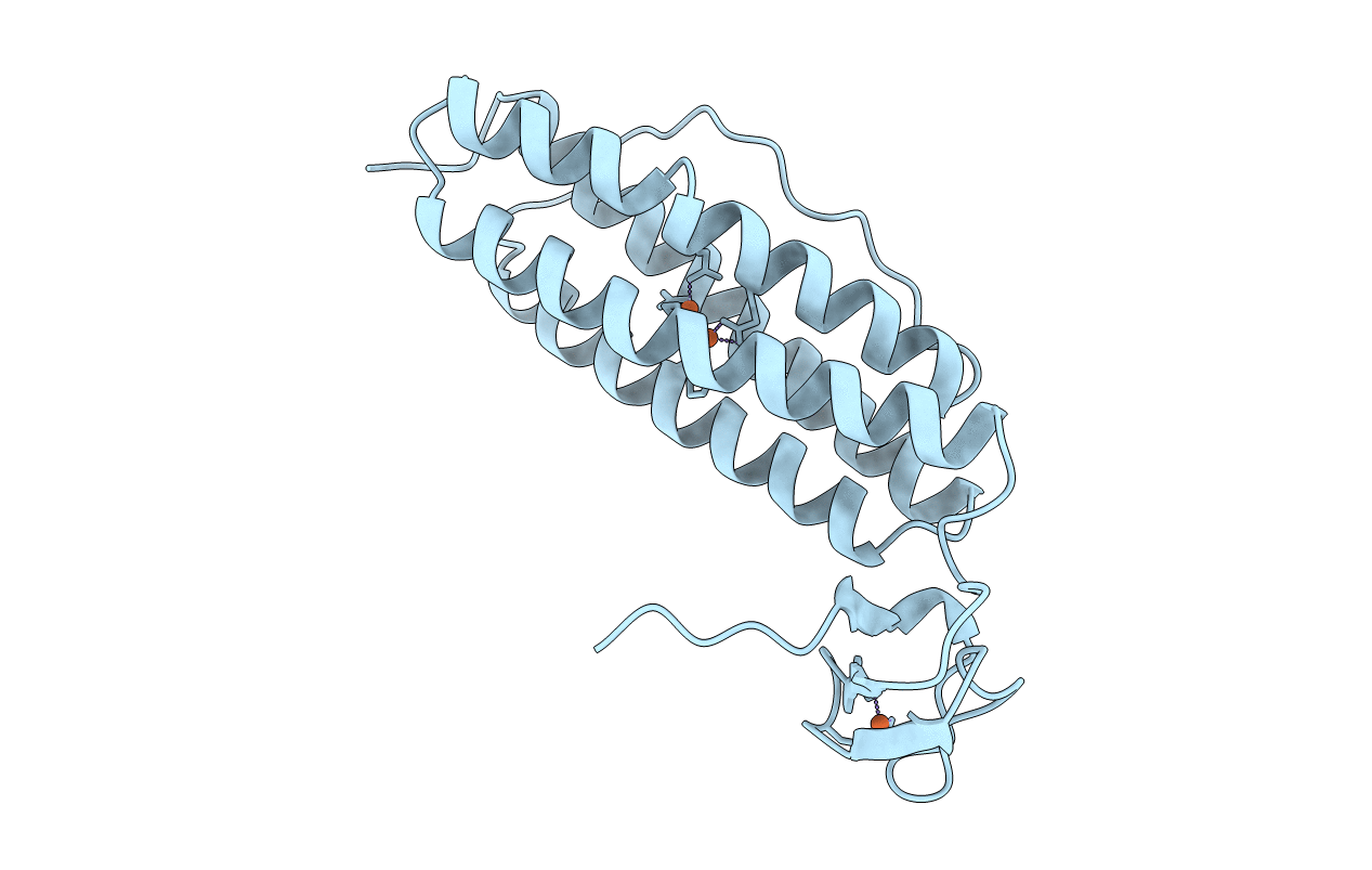
1RYT image
Deposition Date 1996-04-26
Release Date 1997-05-15
Last Version Date 2024-02-14
Entry Detail
PDB ID:
1RYT
Title:
RUBRERYTHRIN
Biological Source:
Method Details:
Experimental Method:
Resolution:
2.10 Å
R-Value Free:
0.25
R-Value Work:
0.18
R-Value Observed:
0.18
Space Group:
I 2 2 2
Macromolecular Entities
Polymer Type:polypeptide(L)
Molecule:RUBRERYTHRIN
Gene (Uniprot):rbr
Chain IDs:A
Chain Length:190
Number of Molecules:1
Biological Source:Desulfovibrio vulgaris subsp. vulgaris str. Hildenborough
Ligand Molecules
Primary Citation
The structure of Desulfovibrio vulgaris rubrerythrin reveals a unique combination of rubredoxin-like FeS4 and ferritin-like diiron domains.
Nat.Struct.Biol. 3 539 546 (1996)
PMID: 8646540 DOI: 10.1038/nsb0696-539

Abstact

We have determined the structure of rubrerythrin, a non-haem iron protein from the anaerobic sulphate-reducing bacterium, Desulfovibrio vulgaris (Hildenborough), by X-ray crystallography. The structure reveals a tetramer of two-domain subunits. Each subunit contains a four-helix bundle surrounding a diiron-oxo site and a C-terminal rubredoxin-like FeS4 domain. The diiron-oxo site contains a larger number of carboxylate ligands and a higher degree of solvent exposure than do those in other diiron-oxo proteins. The four-helix bundle of rubrerythrin closely resembles those of the ferritin and bacterioferritin subunits, suggesting a relationship among these proteins-consistent with the recently demonstrated ferroxidase activity of rubrerythrin.

Legend

Protein

Chemical

Disease

Primary Citation of related structures