Deposition Date
2003-12-17
Release Date
2004-04-13
Last Version Date
2024-03-13
Entry Detail
PDB ID:
1RWR
Keywords:
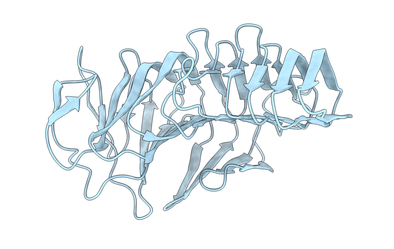
Title:
Crystal structure of filamentous hemagglutinin secretion domain
Biological Source:
Source Organism(s):
Bordetella pertussis (Taxon ID: 520)
Expression System(s):
Method Details:
Experimental Method:
Resolution:
1.72 Å
R-Value Free:
0.18
R-Value Work:
0.14
R-Value Observed:
0.14
Space Group:
C 1 2 1