1RV6 image
Deposition Date 2003-12-12
Release Date 2004-01-20
Last Version Date 2024-11-06
Entry Detail
PDB ID:
1RV6
Title:
Crystal Structure of PlGF in Complex with Domain 2 of VEGFR1
Biological Source:
Source Organism:
Homo sapiens (Taxon ID: 9606)
Host Organism:
Method Details:
Experimental Method:
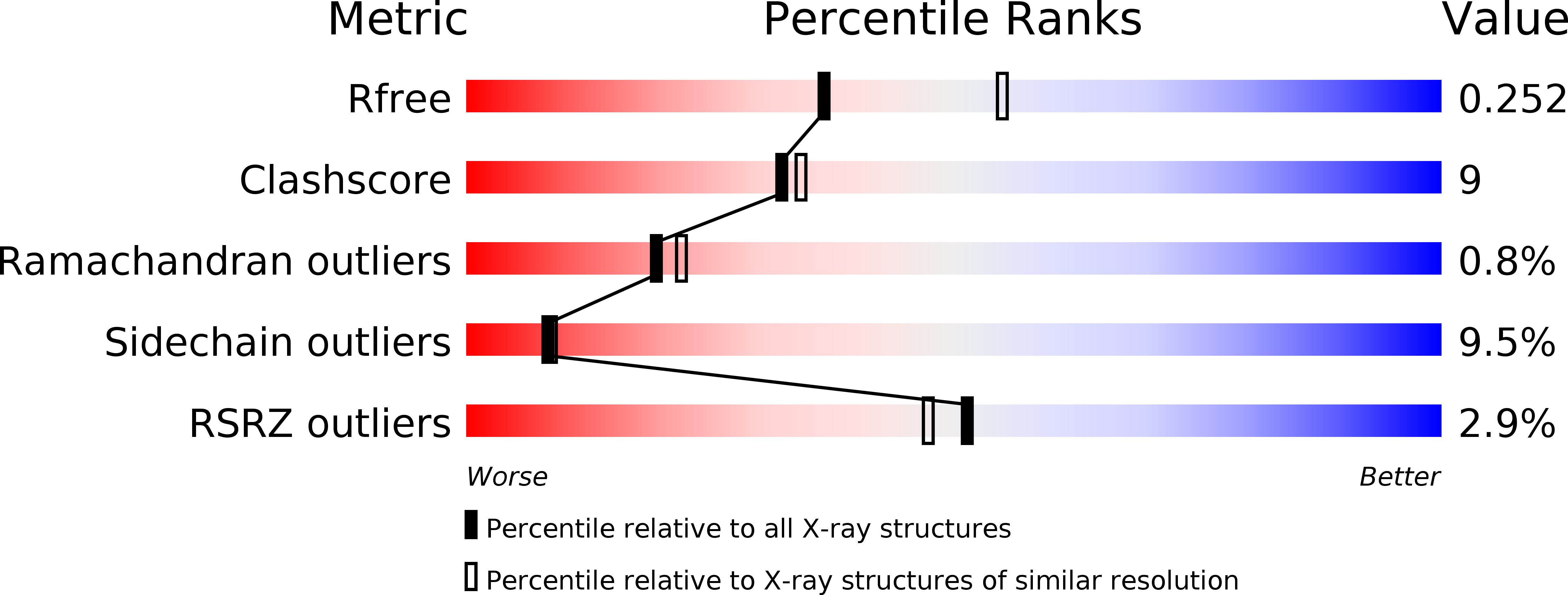
Resolution:
2.45 Å
R-Value Free:
0.26
R-Value Work:
0.19
R-Value Observed:
0.19
Space Group:
P 21 21 21
Macromolecular Entities
Polymer Type:polypeptide(L)
Molecule:placenta growth factor (PlGF)
Gene (Uniprot):PGF
Chain IDs:A (auth: V), B (auth: W)
Chain Length:101
Number of Molecules:2
Biological Source:Homo sapiens
Polymer Type:polypeptide(L)
Molecule:FLT1 protein
Gene (Uniprot):FLT1
Chain IDs:C (auth: X), D (auth: Y)
Chain Length:100
Number of Molecules:2
Biological Source:Homo sapiens
Ligand Molecules
Primary Citation
The crystal structure of placental growth factor in complex with domain 2 of vascular endothelial growth factor receptor-1.
J.Biol.Chem. 279 10382 10388 (2004)
PMID: 14684734 DOI: 10.1074/jbc.M313237200

Abstact

Placental growth factor (PlGF) is a member of the vascular endothelial growth factor (VEGF) family and plays an important role in pathological angiogenic events. PlGF exerts its biological activities through binding to VEGFR1, a receptor tyrosine kinase that consists of seven immunoglobulin-like domains in its extracellular portion. Here we report the crystal structure of PlGF bound to the second immunoglobulin-like domain of VEGFR1 at 2.5 A resolution and compare the complex to the closely related structure of VEGF bound to the same receptor domain. The two growth factors, PlGF and VEGF, share a sequence identity of approximately 50%. Despite this moderate sequence conservation, they bind to the same binding interface of VEGFR1 in a very similar fashion, suggesting that both growth factors could induce very similar if not identical signaling events.

Legend

Protein

Chemical

Disease

Primary Citation of related structures