1RQQ image
Deposition Date 2003-12-06
Release Date 2003-12-30
Last Version Date 2024-10-09
Entry Detail
PDB ID:
1RQQ
Title:
Crystal Structure of the Insulin Receptor Kinase in Complex with the SH2 Domain of APS
Biological Source:
Source Organism:
Homo sapiens (Taxon ID: 9606)
Rattus norvegicus (Taxon ID: 10116)
Host Organism:
Method Details:
Experimental Method:
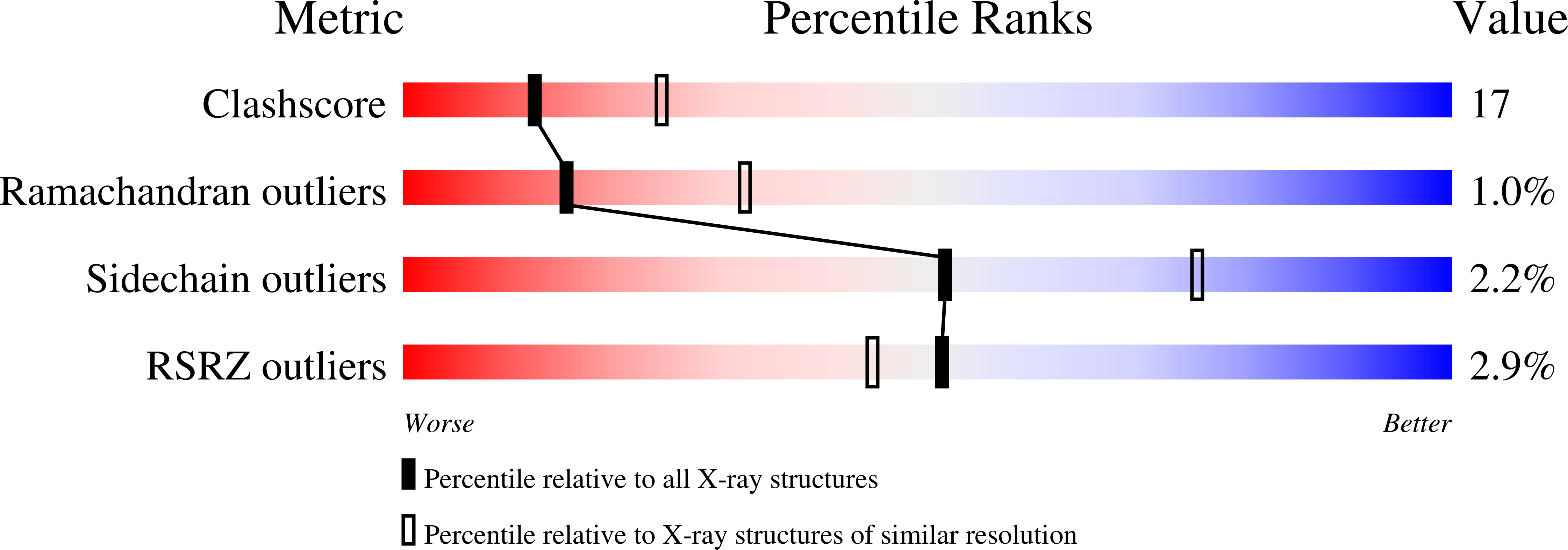
Resolution:
2.60 Å
R-Value Free:
0.27
R-Value Work:
0.22
R-Value Observed:
0.22
Space Group:
P 21 21 21
Macromolecular Entities
Structures with similar UniProt ID
Protein Blast
Polymer Type:polypeptide(L)
Molecule:Insulin receptor
Gene (Uniprot):INSR
Mutagens:C980S, K1251N
Chain IDs:A, B
Chain Length:306
Number of Molecules:2
Biological Source:Homo sapiens
Structures with similar UniProt ID
Protein Blast
Polymer Type:polypeptide(L)
Molecule:adaptor protein APS
Gene (Uniprot):Sh2b2
Chain IDs:C, D
Chain Length:114
Number of Molecules:2
Biological Source:Rattus norvegicus
Modified Residue
Compound ID Chain ID Parent Comp ID Details 2D Image
PTR A TYR O-PHOSPHOTYROSINE
Primary Citation
Structural basis for recruitment of the adaptor protein APS to the activated insulin receptor.
Mol.Cell 12 1379 1389 (2003)
PMID: 14690593 DOI: 10.1016/S1097-2765(03)00487-8

Abstact

The adaptor protein APS is a substrate of the insulin receptor and couples receptor activation with phosphorylation of Cbl to facilitate glucose uptake. The interaction with the activated insulin receptor is mediated by the Src homology 2 (SH2) domain of APS. Here, we present the crystal structure of the APS SH2 domain in complex with the phosphorylated tyrosine kinase domain of the insulin receptor. The structure reveals a novel dimeric configuration of the APS SH2 domain, wherein the C-terminal half of each protomer is structurally divergent from conventional, monomeric SH2 domains. The APS SH2 dimer engages two kinase molecules, with pTyr-1158 of the kinase activation loop bound in the canonical phosphotyrosine binding pocket of the SH2 domain and a second phosphotyrosine, pTyr-1162, coordinated by two lysine residues in beta strand D. This structure provides a molecular visualization of one of the initial downstream recruitment events following insulin activation of its dimeric receptor.

Legend

Protein

Chemical

Disease

Primary Citation of related structures