Deposition Date
2003-12-03
Release Date
2004-02-17
Last Version Date
2024-11-20
Entry Detail
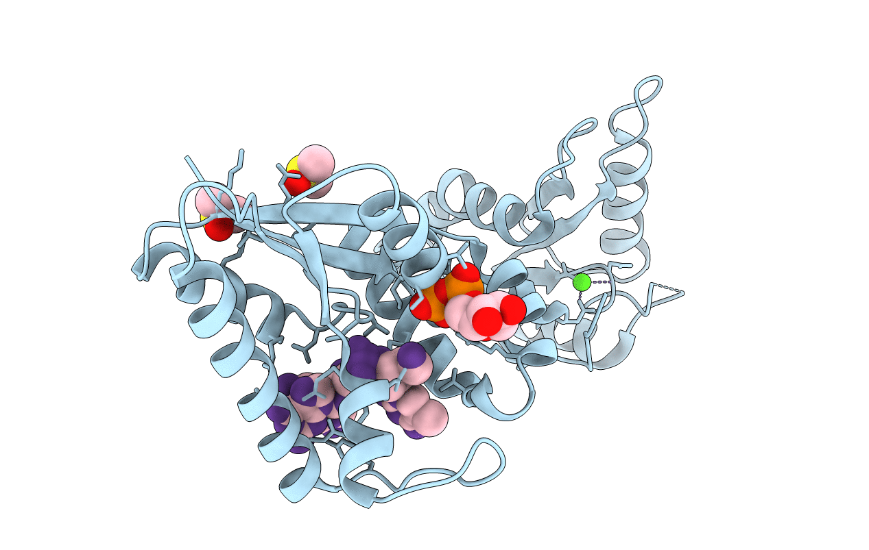
Biological Source:
Source Organism(s):
Enterobacteria phage T4 (Taxon ID: 10665)
Expression System(s):
Method Details:
Experimental Method:
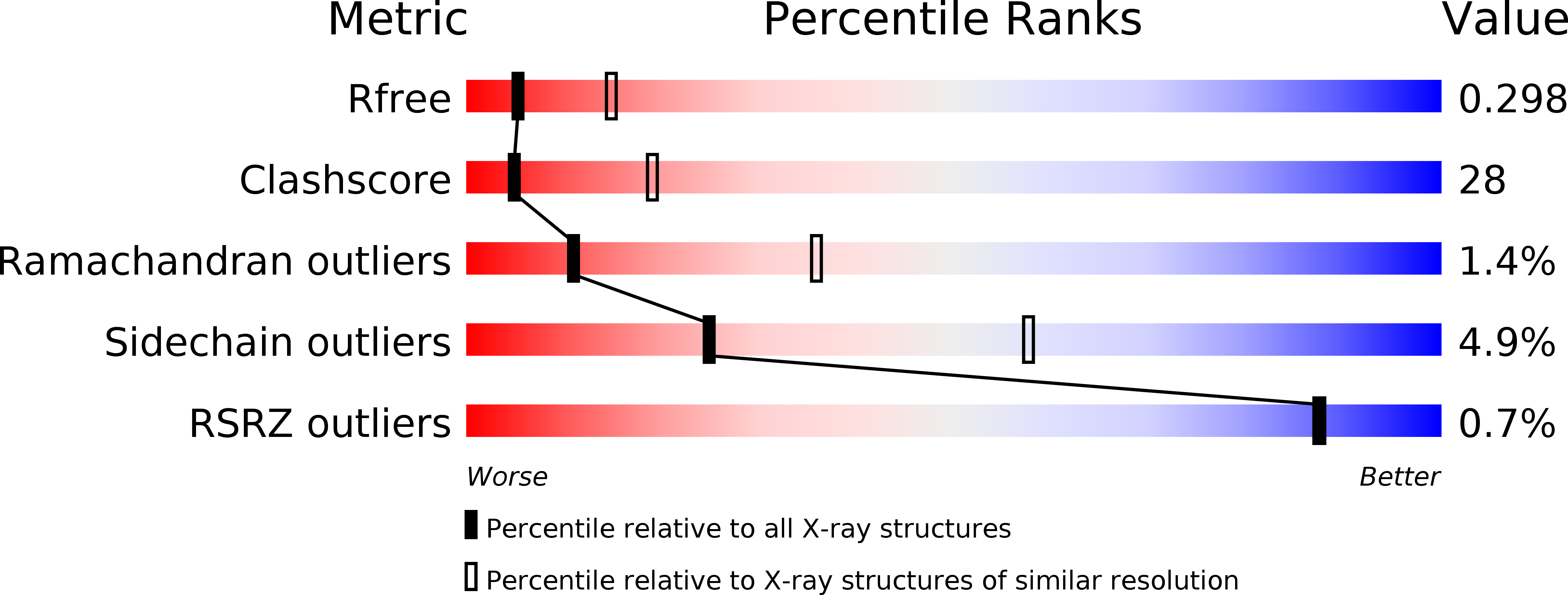
Resolution:
2.90 Å
R-Value Free:
0.30
R-Value Work:
0.23
R-Value Observed:
0.23
Space Group:
I 2 2 2