1RPN image
Deposition Date 2003-12-03
Release Date 2004-02-24
Last Version Date 2023-08-23
Entry Detail
PDB ID:
1RPN
Keywords:
Title:
Crystal Structure of GDP-D-mannose 4,6-dehydratase in complexes with GDP and NADPH
Biological Source:
Source Organism:
Host Organism:
Method Details:
Experimental Method:
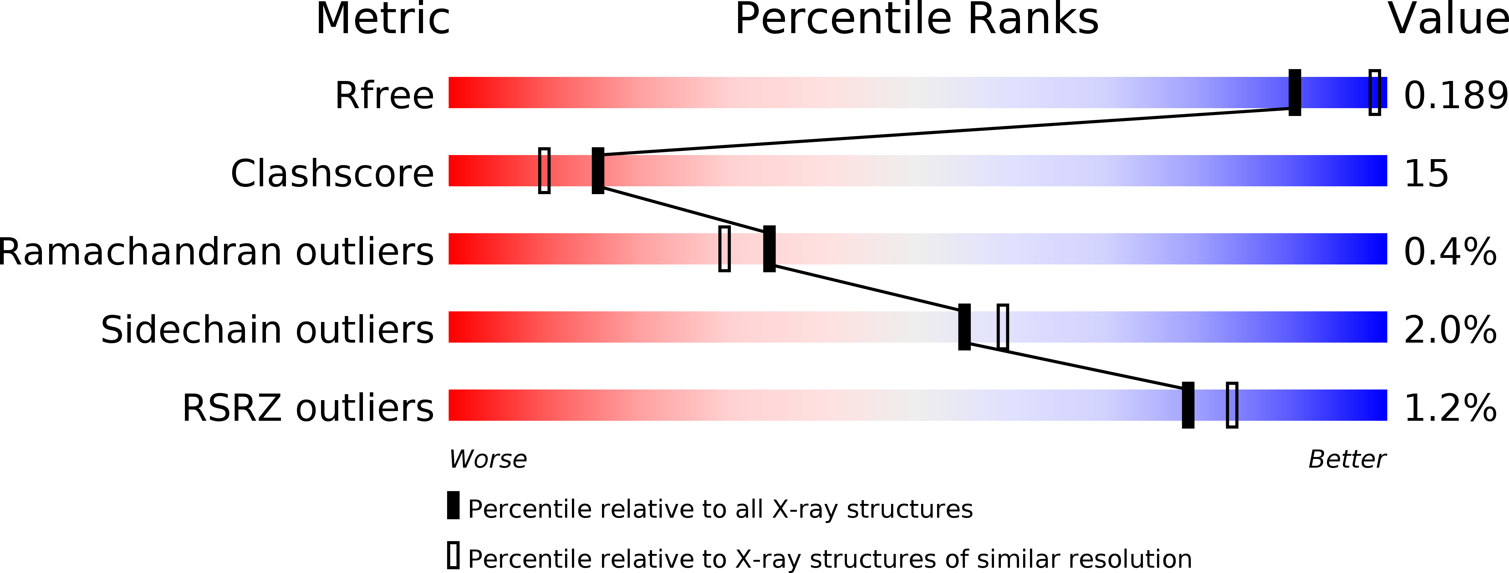
Resolution:
2.15 Å
R-Value Free:
0.19
R-Value Work:
0.17
R-Value Observed:
0.17
Space Group:
P 32 2 1
Macromolecular Entities
Structures with similar UniProt ID
Protein Blast
Polymer Type:polypeptide(L)
Molecule:GDP-mannose 4,6-dehydratase
Gene (Uniprot):gmd
Chain IDs:A, B, C, D
Chain Length:335
Number of Molecules:4
Biological Source:Pseudomonas aeruginosa
Primary Citation
Crystal structure of a tetrameric GDP-D-mannose 4,6-dehydratase from a bacterial GDP-D-rhamnose biosynthetic pathway.
Protein Sci. 13 529 539 (2004)
PMID: 14739333 DOI: 10.1110/ps.03393904

Abstact

d-Rhamnose is a rare 6-deoxy monosaccharide primarily found in the lipopolysaccharide of pathogenic bacteria, where it is involved in host-bacterium interactions and the establishment of infection. The biosynthesis of d-rhamnose proceeds through the conversion of GDP-d-mannose by GDP-d-mannose 4,6-dehydratase (GMD) to GDP-4-keto-6-deoxymannose, which is subsequently reduced to GDP-d-rhamnose by a reductase. We have determined the crystal structure of GMD from Pseudomonas aeruginosa in complex with NADPH and GDP. GMD belongs to the NDP-sugar modifying subfamily of the short-chain dehydrogenase/reductase (SDR) enzymes, all of which exhibit bidomain structures and a conserved catalytic triad (Tyr-XXX-Lys and Ser/Thr). Although most members of this enzyme subfamily display homodimeric structures, this bacterial GMD forms a tetramer in the same fashion as the plant MUR1 from Arabidopsis thaliana. The cofactor binding sites are adjoined across the tetramer interface, which brings the adenosyl phosphate moieties of the adjacent NADPH molecules to within 7 A of each other. A short peptide segment (Arg35-Arg43) stretches into the neighboring monomer, making not only protein-protein interactions but also hydrogen bonding interactions with the neighboring cofactor. The interface hydrogen bonds made by the Arg35-Arg43 segment are generally conserved in GMD and MUR1, and the interacting residues are highly conserved among the sequences of bacterial and eukaryotic GMDs. Outside of the Arg35-Arg43 segment, residues involved in tetrameric contacts are also quite conserved across different species. These observations suggest that a tetramer is the preferred, and perhaps functionally relevant, oligomeric state for most bacterial and eukaryotic GMDs.

Legend

Protein

Chemical

Disease

Primary Citation of related structures
Feedback Form
Name
Email
Institute
Feedback