1RPK image
Deposition Date 2003-12-03
Release Date 2005-06-07
Last Version Date 2023-08-23
Entry Detail
PDB ID:
1RPK
Keywords:
Title:
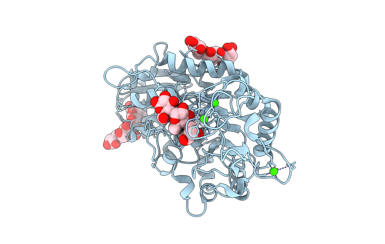
Crystal structure of barley alpha-amylase isozyme 1 (amy1) in complex with acarbose
Biological Source:
Source Organism:
Hordeum vulgare (Taxon ID: 4513)
Host Organism:
Method Details:
Experimental Method:
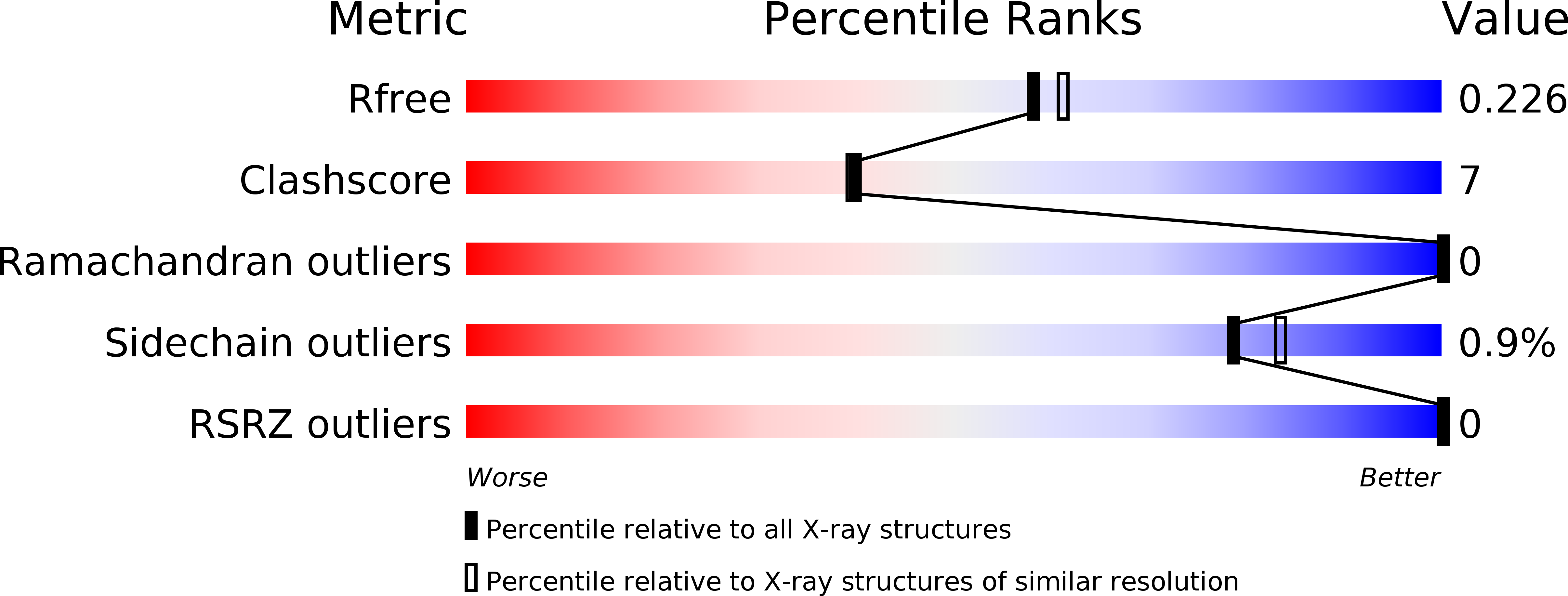
Resolution:
2.00 Å
R-Value Free:
0.23
R-Value Work:
0.18
R-Value Observed:
0.18
Space Group:
P 21 21 2
Macromolecular Entities
Structures with similar UniProt ID
Protein Blast
Polymer Type:polypeptide(L)
Molecule:Alpha-amylase type 1 isozyme
Gene (Uniprot):AMY1.1
Chain IDs:A
Chain Length:405
Number of Molecules:1
Biological Source:Hordeum vulgare
Ligand Molecules
Primary Citation
Oligosaccharide Binding to Barley {alpha}-Amylase 1
J.Biol.Chem. 280 32968 32978 (2005)
PMID: 16030022 DOI: 10.1074/jbc.M505515200

Abstact

Enzymatic subsite mapping earlier predicted 10 binding subsites in the active site substrate binding cleft of barley alpha-amylase isozymes. The three-dimensional structures of the oligosaccharide complexes with barley alpha-amylase isozyme 1 (AMY1) described here give for the first time a thorough insight into the substrate binding by describing residues defining 9 subsites, namely -7 through +2. These structures support that the pseudotetrasaccharide inhibitor acarbose is hydrolyzed by the active enzymes. Moreover, sugar binding was observed to the starch granule-binding site previously determined in barley alpha-amylase isozyme 2 (AMY2), and the sugar binding modes are compared between the two isozymes. The "sugar tongs" surface binding site discovered in the AMY1-thio-DP4 complex is confirmed in the present work. A site that putatively serves as an entrance for the substrate to the active site was proposed at the glycone part of the binding cleft, and the crystal structures of the catalytic nucleophile mutant (AMY1D180A) complexed with acarbose and maltoheptaose, respectively, suggest an additional role for the nucleophile in the stabilization of the Michaelis complex. Furthermore, probable roles are outlined for the surface binding sites. Our data support a model in which the two surface sites in AMY1 can interact with amylose chains in their naturally folded form. Because of the specificities of these two sites, they may locate/orient the enzyme in order to facilitate access to the active site for polysaccharide chains. Moreover, the sugar tongs surface site could also perform the unraveling of amylose chains, with the aid of Tyr-380 acting as "molecular tweezers."

Legend

Protein

Chemical

Disease

Primary Citation of related structures
Feedback Form
Name
Email
Institute
Feedback