Deposition Date
1997-03-24
Release Date
1997-10-15
Last Version Date
2024-02-14
Entry Detail
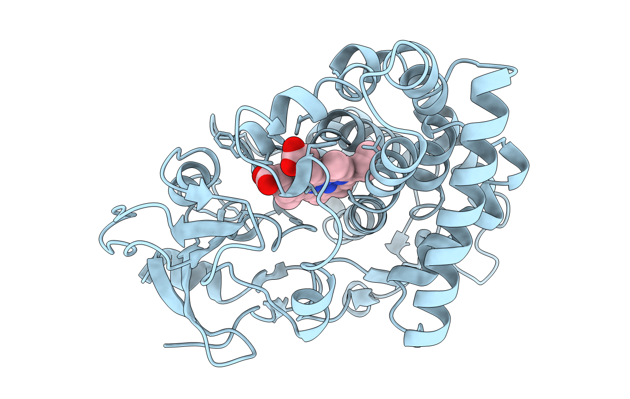
PDB ID:
1ROM
Keywords:
Title:
CRYSTAL STRUCTURE OF NITRIC REDUCTASE FROM DENITRIFYING FUNGUS FUSARIUM OXYSPORUM
Biological Source:
Source Organism(s):
Fusarium oxysporum (Taxon ID: 5507)
Method Details:
Experimental Method:
Resolution:
2.00 Å
R-Value Free:
0.26
R-Value Work:
0.19
R-Value Observed:
0.19
Space Group:
P 21 21 21